
VQ-Wave: Rethinking Lung MRI through Physics-Driven "Chaos" Training
VQ-Wave: A physics-driven spatio-temporal deep learning approach for non-contrast-enhanced lung ventilation and perfusion MRI
VQ-Wave is a physics-driven spatio-temporal deep learning framework designed for non-contrast-enhanced lung functional MRI. It utilizes an Inception-based neural network trained on synthetic stochastic signals to estimate ventilation and perfusion parameters, achieving state-of-the-art robustness against physiological non-stationarity.
TL;DR
Quantifying lung function without contrast agents usually fails when patients don't breathe like a metronome. VQ-Wave breaks this limitation using a spatio-temporal Inception network trained on synthetic "chaotic" signals. It cuts MRI scan times by 66% (from 45s to 15s) while maintaining quantitative stability, even in pediatric patients with irregular physiology.
Problem & Motivation: The "Stationarity" Trap
Current non-contrast lung MRI relies on Fourier Decomposition or Matrix Pencil (MP) methods. These techniques view the body as a collection of perfect oscillators. They assume your heart beats and lungs move with mathematical regularity.
The Reality Check: Patients—especially children with Cystic Fibrosis (CF)—don't breathe perfectly. They sigh, they cough, and their heart rates drift. To a traditional algorithm, this non-stationarity causes spectral leakage. Imagine a radio station slightly off-frequency; the signal becomes static. In MRI, this results in signal dropouts, noise-filled perfusion maps, and unreliable clinical data.
Methodology: Learning from Synthetic Chaos
The authors' masterstroke wasn't just the architecture, but the training data. Since "ground truth" for a single voxel's ventilation in a living person is impossible to measure, they built a Stochastic Physiological Simulator.
1. The Physics-Driven Generator
The generator creates 3x3 voxel patches with infinite variations of:
- Frequency drifts (shifting breath rates).
- Amplitude modulations (deep sighs vs. shallow breaths).
- Non-linear motion (using the Lujan model).
- Heavy noise profiles.
2. The Inception-SE Architecture
The network (VQ-Wave) uses 1D Inception blocks with varying kernels (from to ). This allows it to "see" both fast cardiac pulses and slow respiratory trends simultaneously.
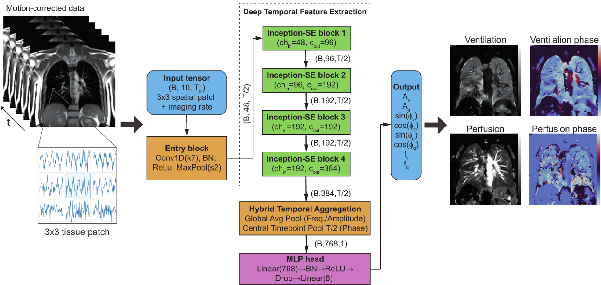 Figure: The VQ-Wave architecture combines multi-scale temporal kernels with Squeeze-and-Excitation attention to filter noise locally.
Experiments & Results: Robustness Under Fire
The researchers tested VQ-Wave against the industry-standard Matrix Pencil (MP) method across three brutal benchmarks: noise, instability, and scan length.
Superior Denoising
Even at high noise levels (), VQ-Wave maintained sharp vascular edges while MP maps became "grainy" and non-diagnostic.
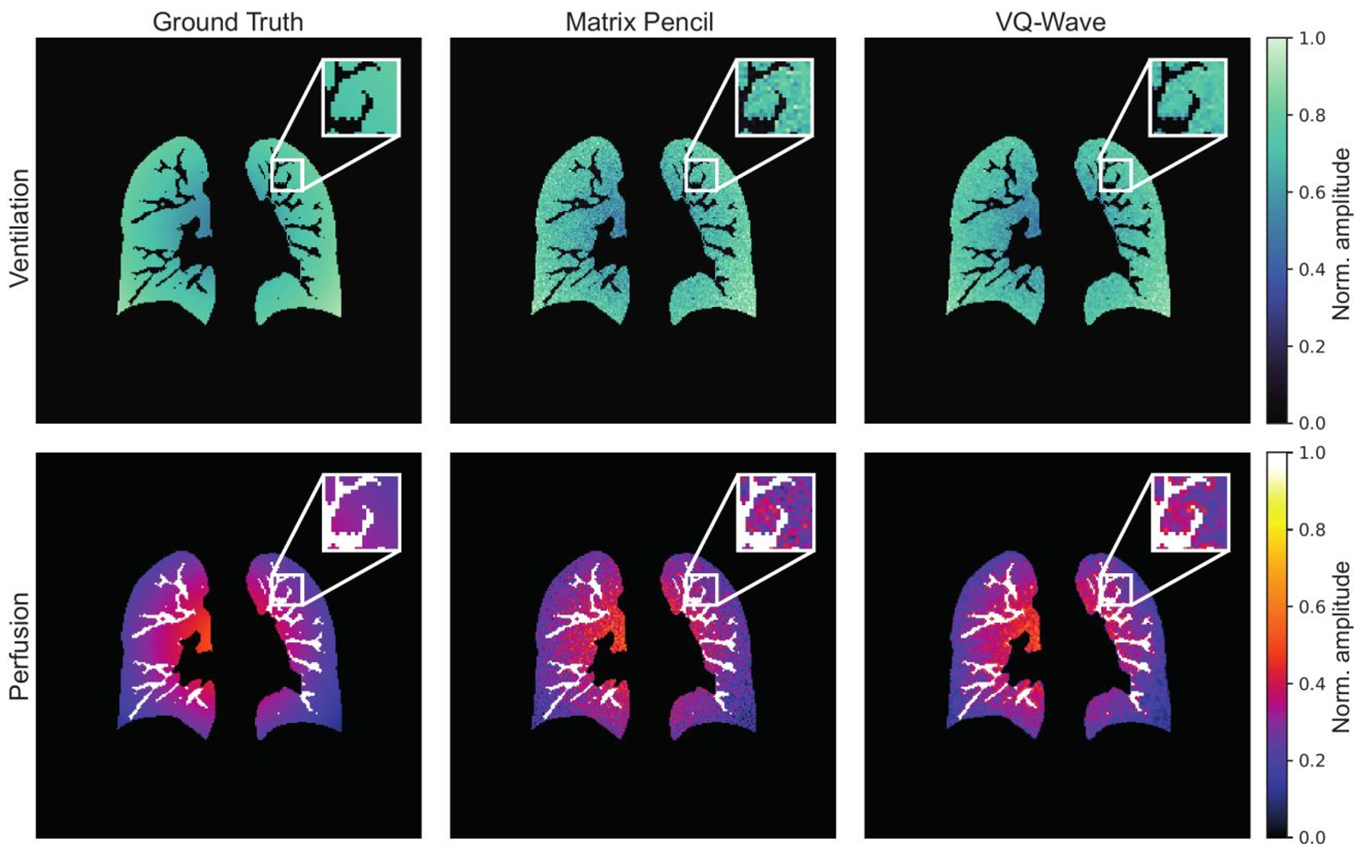 Figure: VQ-Wave preserves anatomical boundaries (vessels) significantly better than MP decomposition under high-noise conditions.
The 15-Second Milestone
The most impactful result is scan time reduction. Traditional methods require at least 80-140 images to resolve frequencies. VQ-Wave achieved stable, diagnostic results with just 40 images (15 seconds).
In clinical tests on a 14-year-old CF patient, VQ-Wave's perfusion maps remained coherent when the scan was truncated, whereas the MP method's data essentially collapsed into noise (60% volatility vs. <12% for VQ-Wave).
Critical Analysis & Conclusion
VQ-Wave marks a transition from "Rigid Signal Processing" to "Adaptive Neural Filtering."
Why it works:
- Inductive Bias: By using a 3x3 spatial context, the network acts as a smart arbiter—it knows when to smooth noise and when to preserve a sharp vessel boundary.
- Domain Agnosticism: Because it's trained on physics models rather than specific images, it can theoretically work on any MRI scanner (0.55T to 3T) without retraining.
Limitations:
While the synthetic simulator is powerful, it may not yet capture every nuance of severe pulmonary disease (like extreme partial volume effects). However, the spatial overlap between VQ-Wave and the gold-standard MP (Dice coefficient of 0.96 for ventilation) proves it is clinically ready for larger trials.
Final Takeaway: This work proves that we don't need longer scans; we need smarter filters that embrace the "chaos" of human physiology.