
Regional Multi-View Learning: Elevating Ultrasound Diagnosis with MR Intelligence
310_Regional Multi-View Learning for Cardiac Motion An
The paper introduces an automated diagnostic pipeline for identifying Dilated Cardiomyopathy (DCM) using 3D ultrasound (US) data, enhanced by a multimodal cardiac motion atlas. It leverages regional multi-view learning (MLDA and MvLapSVM) to incorporate high-fidelity motion descriptors from magnetic resonance (MR) imaging during training to improve US-based classification, achieving a top accuracy of 94.32%.
TL;DR
Researchers have developed a sophisticated "Regional Multi-View" diagnostic pipeline that allows low-cost 3D Ultrasound (US) to reach the diagnostic accuracy of High-Field MR. By training on a multimodal cardiac motion atlas and using advanced manifold learning (MvLapSVM), the system identifies Dilated Cardiomyopathy (DCM) with 94.32% accuracy, proving that the "physics" of heart motion learned from MR can be effectively transferred to enhance US-based clinical workflows.
The Motivation: High-Fidelity Motion vs. Low-Cost Access
The clinical dilemma in cardiology is a trade-off between precision and accessibility.
- 3D Tagged MR is the "Gold Standard" for Myocardial Wall Motion Analysis (MWMA), providing exquisite detail on how heart muscles stretch and twist. But it is expensive, slow, and unsuitable for many patients (e.g., those with pacemakers).
- Ultrasound (US) is the clinical workhorse—cheap and portable—but it is plagued by "acoustic shadowing" and noise, making localized motion analysis prone to error.
The authors ask: Can we use a database of simultaneous MR/US captures to "teach" an ultrasound-only system what high-quality cardiac motion looks like?
Methodology: Mining Multi-View Manifolds
The core innovation lies in the transition from global analysis to Regional Multi-View Learning.
1. The Multimodal Spatiotemporal Atlas
The pipeline begins by normalizing all heart geometries into a single reference space. Using a B-spline Free-Form Deformation (FFD) registration, the researchers estimate 3D displacements across the cardiac cycle for both MR and US views.
2. The Multi-View Learning Engine
Instead of just concatenating data (which leads to overfitting), the authors utilize Multi-view Laplacian Support Vector Machines (MvLapSVM). This algorithm assumes that while MR and US look different, they describe the same underlying "manifold" of cardiac mechanics. The objective function minimizes the disagreement between the MR-learner and the US-learner.
3. Regional Sensitivity
The heart isn't a uniform pump. The authors divided the left ventricle into 17 American Heart Association (AHA) regions. They found that localized motion in the basal and mid-anterior segments provided the most discriminative information.
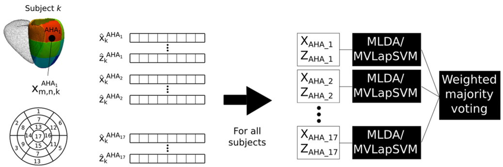 Figure: The Regional Multi-View strategy, showing how data is partitioned by AHA segments before classification.
Experimental Results: Beyond Ejection Fraction
The results validate the hypothesis that multimodal training is superior to single-modality approaches:
| Method | Balanced Accuracy (BACC) |
|---|---|
| Standard EF (Clinical Baseline) | 82.5% |
| US-Only (Single Modality) | 87.3% |
| Regional MvLapSVM (Proposed) | 94.3% |
The "misclassification analysis" revealed that the model struggles only when clinical parameters (like Ejection Fraction) are borderline—matching the difficulty even human experts face in early-stage DCM.
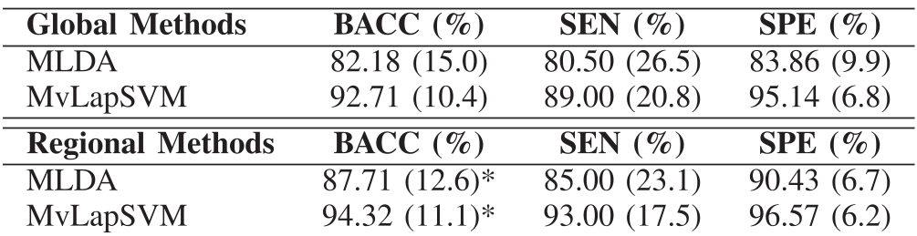 Table: Comparison of Global vs. Regional methods showing the significant accuracy boost from regional analysis.
Critical Analysis & Professional Insights
Why it works: The regional approach acts as an Inductive Bias. By forcing the model to look at specific anatomical segments, the researchers reduced the "Curse of Dimensionality" (too many features, too few patients). The incorporation of MR info during training acts as a regularizer, preventing the US model from being fooled by noise patterns unique to ultrasound.
Limitations: The dataset, while multi-center, is relatively small (19 DCM, 50 Healthy). Future validation on larger, more heterogeneous "real-world" cohorts is needed. Furthermore, the pipeline currently ignores non-imaging data (age, genetics) which could further boost performance.
Conclusion
This paper marks a shift in medical AI from "single-modality" black boxes to "physically-informed" multimodal pipelines. By embedding this logic into US scanners, we could soon see "Standard" ultrasound machines providing "MR-quality" diagnostic insights at the bedside.