
Beyond Tissue Density: Deciphering Gender from Brain Water Diffusion with 3D-CNNs
A 3D-CNN Classifier for Gender Discrimination from Diffusion Tensor Imaging of Human Brain
This paper introduces a multi-channel 3D-CNN classifier designed for gender discrimination using Diffusion Tensor Imaging (DTI). By mapping DTI's directional water molecule movement into distinct input channels, the model achieves a gender classification accuracy of 85.1%, comparable to models using standard T1 and T2 weighted structural brain images.
TL;DR
Researchers at Kochi University of Technology have developed a novel multi-channel 3D-CNN that performs gender classification by "reading" how water molecules move through the human brain. By repurposing the 16 directional tensors of Diffusion Tensor Imaging (DTI) as separate input channels, the model achieves an 85.1% accuracy, proving that molecular diffusion patterns contain significant demographic signals traditionally extracted from structural T1-weighted images.
The Structural Ceiling and the Diffusion Insight
In the realm of Neuroimaging, T1-weighted images are the gold standard because they provide clear anatomical maps of gray and white matter. However, T1 images essentially represent a "still life" of brain density.
The authors argue that we are missing a dimension: Diffusion Tensor Imaging (DTI). DTI measures the movement of water molecules, providing a proxy for the brain's structural connectivity (its "wiring"). The challenge? DTI data is not a simple scalar value (like brightness) but a tensor representing movement in multiple directions. Standard 3D-CNNs, designed for single-channel volumes, cannot ingest this complexity directly.
Methodology: Mapping Tensors to Channels
The core innovation lies in the data pipeline. Instead of trying to collapse the diffusion information, the researchers treated each of the 16 diffusion directions captured in the IXI Dataset as a distinct channel.
The 3D-CNN Architecture
The model follows a rigorous deep learning structure:
- Feature Extraction: 5 layers of 3D Convolution, followed by Batch Normalization and ReLU activation.
- Optimization: Adam optimizer (lr=10⁻³) was selected after preliminary testing for its superior convergence.
- Input Strategy: For T1/T2 images, the input is
(Voxel_H, Voxel_W, Voxel_D, 1). For DTI, it scales to(Voxel_H, Voxel_W, Voxel_D, 16).
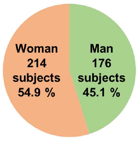 Figure 1: The proposed 7-layer 3D-CNN structure designed for processing multi-modal brain MRI volumes.
Experiments and Competitive Benchmarking
The researchers conducted a comparative study between T1, T2, and DTI using 5-fold cross-validation on a dataset of 390 subjects.
The Peripheral Feature Bias
An intriguing finding emerged during preprocessing. When the images were not "skull-stripped" (keeping fat and external tissues), T1-weighted images reached a near-perfect 98.6% accuracy. However, when using FSL to extract only the internal brain tissue, the accuracy dropped to 83.3%.
This suggests that CNNs might be "cheating" by looking at head shape or subcutaneous fat rather than actual brain structure. In contrast, the DTI results (85.1%) remained robust, indicating that the internal diffusion patterns are genuinely discriminative.
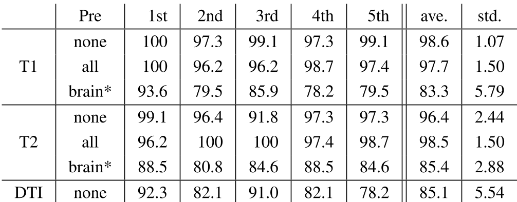 Table 2: Accuracy comparison across different MRI sequences and preprocessing levels.
Critical Analysis & Professional Takeaways
While the T1 results without preprocessing are higher, the DTI approach is arguably more scientifically valuable. DTI captures microstructural integrity that is invisible to T1 scans.
Key Technical Lessons:
- Inductive Bias: Using directional DTI as channels allows the network to learn spatial relationships between diffusion vectors.
- Dataset Sensitivity: The accuracy drop after brain extraction highlights the importance of rigorous preprocessing in medical AI—models often pick up on "non-brain" signals to classify biological traits.
- Future Utility: The successful classification of gender using DTI paves the way for detecting harder-to-spot signals, such as early-stage Alzheimer’s or mental health conditions, where macro-structural changes are absent but diffusion patterns are already altered.
Conclusion
The study successfully transitions DTI from a clinical diagnostic tool to a deep learning-compatible feature set. While T1 scans remain dominant for now, the multi-channel 3D-CNN approach provides a blueprint for leveraging the "movement of water" to understand the human brain.