
3D CNN: Reimagining EEG Signals as Spatio-Temporal Volumes for Superior Emotion Recognition
A 3D Convolutional Neural Network for Emotion Recognition based on EEG Signals
This paper presents a 3D Convolutional Neural Network (3D CNN) framework designed for EEG-based emotion recognition. By transforming raw EEG signals into a 2D electrode topological structure and stacking them along the temporal dimension, the model effectively extracts spatio-temporal features, achieving SOTA accuracies of 96.61% and 97.52% for two-class classification on the DEAP and AMIGOS datasets, respectively.
TL;DR
Decoding human emotions from brainwaves (EEG) has long struggled with the loss of spatial context and temporal dynamics. This paper introduces a 3D Convolutional Neural Network that treats EEG data not as simple time-series, but as a "video" of brain activity. By mapping 32-channel signals into a 9x9 topological grid and applying 3D kernels, the authors achieved an unprecedented 96.61% accuracy on the DEAP dataset, setting a new benchmark for affective computing.
Problem & Motivation: The Gap in EEG Feature Engineering
In the realm of Human-Machine Interaction, EEG signals are the "Gold Standard" because they cannot be subjectively faked like facial expressions. However, most researchers face a dilemma:
- Traditional ML: Relies on hand-crafted features (e.g., Power Spectral Density), which are only as good as the designer's domain knowledge.
- 2D Deep Learning: Often treats channels as independent pixels or collapses the spatial layout of the brain, losing the critical information of where the signal comes from on the scalp.
The authors' insight was simple yet powerful: To truly understand an emotion, we must look at how electrical activity moves across the brain's surface over time. This requires 3D feature extraction.
Methodology: Mapping the Scalp to a Volume
The core innovation lies in the transformation of raw data into a format suitable for 3D convolution.
1. Baseline Pre-processing
Emotional signals are relative. The authors cut 3 seconds of "baseline" (no stimulus) signals, averaged them, and subtracted this mean from the experimental data. This "baseline removal" acts as a form of physiological normalization, which has been shown to increase accuracy by up to 30%.
2. Electrode Topological Relocation
Standard EEG datasets provide signals in a flat list (Channel 1, 2, 3...). The authors used the International 10-20 system to map these channels onto a 9x9 matrix.
- Spatial Context: Channels near each other on the scalp are now neighbors in the matrix.
- Temporal Depth: 128 consecutive time points are stacked, creating a 9x9x128 volume.
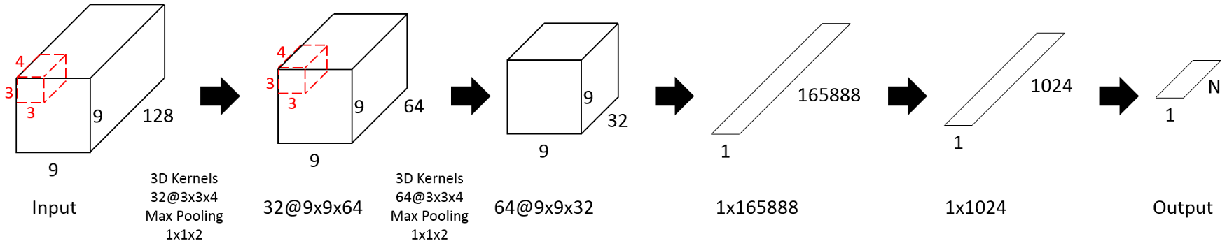 Above: The 3D CNN architecture uses 3x3x4 kernels to extract joint spatial-temporal features, followed by Max-Pooling to consolidate temporal information.
Experiments & Results: Crushing the Baselines
The model was validated on two major benchmarks: DEAP and AMIGOS.
DEAP Dataset Performance
The 3D CNN model achieved over 96% accuracy for binary classification (Arousal/Valence) and 93.53% for the complex four-class task (LALV, HALV, etc.).
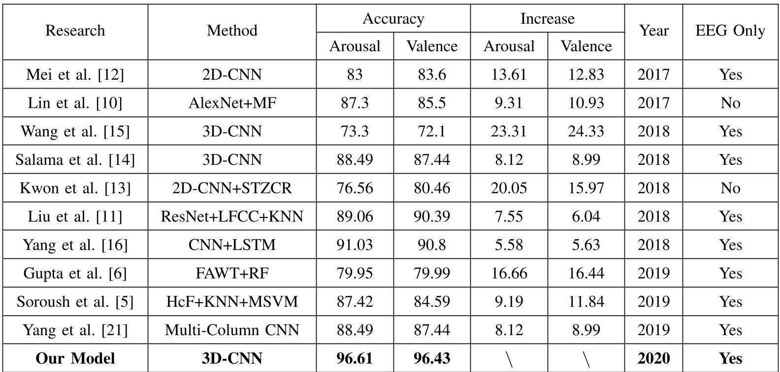 Note the "Increase" column: Compared to previous 3D-CNN and 2D-CNN models, this approach yields an 8% to 23% improvement.
AMIGOS Dataset Performance
The results were even more striking on the newer AMIGOS dataset, reaching 97.52% for Arousal classification, demonstrating that the architecture is robust across different recording setups and participant groups.
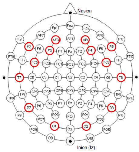 Example of the 2D Electrode Topological Structure (9x9) used for mapping DEAP's 32 channels.
Critical Analysis & Conclusion
The success of this 3D CNN model stems from its Inter-disciplinary Inductive Bias: it respects the physical reality of the human brain (spatial topology) and the nature of electrical signals (temporality).
Key Takeaways:
- Generalization: The same architecture worked across DEAP and AMIGOS without parameter changes.
- Simplicity vs. Power: By getting the data representation right (9x9xT), the model doesn't need to be excessively deep to outperform complex hybrid models (like CNN+LSTM).
Limitations & Future Work: While highly accurate, 3D CNNs are often criticized as "Black Boxes." The authors acknowledge the need to improve explainability—identifying which specific brain regions or frequency bands contribute most to a specific emotion. Future extensions could see this model applied to Motor Imagery (controlling prosthetics) or Dementia Stage Classification, where spatio-temporal patterns are equally critical.