
Decoding the Intracellular Highway: ML-Powered 3D Tracking of Organelle Movement
3D Nanoscale Tracking Data Analysis for Intracellular Organelle Movement using Machine Learning Approach
The paper introduces a machine learning framework for the 3D nanoscale tracking of intracellular organelles. By utilizing a Support Vector Machine (SVM) within an Error-Correcting Output Codes (ECOC) model, the researchers successfully classified vesicle transport transitions between actin filaments and microtubules with an accuracy of 88.4%.
TL;DR
Researchers from the University of Tokyo have developed a machine learning-based approach to solve a long-standing mystery in cell biology: identifying how vesicles switch between different "biological tracks" (actin filaments and microtubules). By analyzing 3D nanoscale trajectory data with a quadratic SVM, they achieved 88.4% accuracy in classifying transport transitions, providing a blueprint for automated analysis of complex intracellular dynamics.
Background & Motivation: The Complexity of the Cytoplasm
Inside a living cell, nutrients and signals are packaged into vesicles and transported like cargo on a massive, intertwined highway system composed of actin filaments and microtubules.
While modern super-resolution microscopy allows us to see these movements at the nanoscale, the data is notoriously difficult to interpret. Current numerical methods can tell that a vesicle is moving, but they struggle to identify which track the vesicle is on at any any given moment because the network is so dense and "tangled." This paper addresses the missing link: identifying the specific cytoskeleton interactions from raw 3D coordinate data.
Methodology: From Physics to Features
The authors framed the problem as a multi-class classification task. They identified four types of transfers based on the biological "switch" occurring:
- A to A: Actin to Actin
- A to M: Actin to Microtubule
- M to A: Microtubule to Actin
- M to M: Microtubule to Microtubule
Feature Engineering
Rather than using raw coordinates, the authors extracted "Physically Informed Features":
- Velocity Difference (): Vesicles move slower on actin than on microtubules.
- Run Length Difference (): Microtubules typically allow for longer continuous runs.
- Transfer Angle (): The geometry of the branching network.
- Transfer Time (): The pause duration during a track switch.
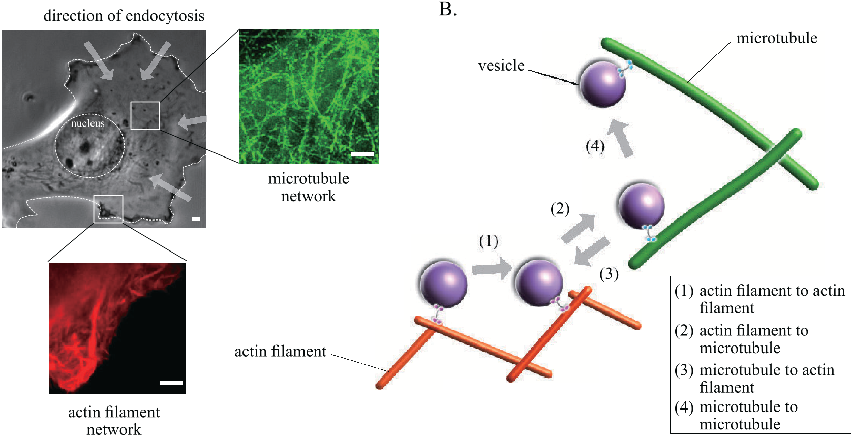 Fig 1. Schematic of the intracellular network and the four classes of vesicle transfer (Source: Lee et al.)
Experimental Results
Using 146 labeled observations from breast cancer cell imaging, the team applied Sequential Feature Selection. Interestingly, they found that Velocity Difference and Run Length were the only two essential features needed for high-accuracy classification.
Using a Quadratic Support Vector Machine (SVM) within an Error-Correcting Output Codes (ECOC) framework, the model achieved an impressive 88.4% accuracy.
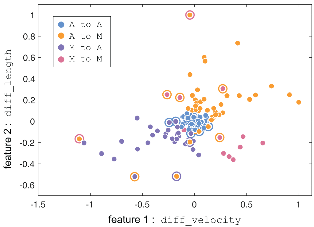 Fig 2. Prediction results: The "diff_velocity" vs "diff_length" feature space successfully clusters the different transfer types.
Critical Insight & Future Outlook
The success of this approach lies in its simplicity. By reducing complex 3D trajectories into physically meaningful parameters (velocity and length difference), the authors avoided the "black box" nature of deep learning while still achieving SOTA-level classification accuracy for this specific niche.
Limitations: The current dataset is relatively small (146 observations). For more heterogeneous cell types or drug-response studies, more robust data and perhaps unsupervised methods might be required to discover "hidden" transport states.
Takeaway: This study proves that ML isn't just for big data; in the realm of nanoscopy, even small-scale "smart" feature engineering can unlock biological insights that were previously hidden in the noise of the cytoplasm.