
Calcium's Hidden Hand: How Functional Reentry Destabilizes Cardiac Tissue
7561_Functional reentry's influence on intracellular calcium in the LRd membrane equations.
This paper investigates the interplay between functional reentry, transmembrane potential, and intracellular calcium () handling using the Luo-Rudy dynamic (LRd) membrane equations. It identifies that the rapid rate of reentry promotes spontaneous sarcoplasmic reticulum release (SCR), particularly within the functional center, leading to wavefront fractionation and reentrant instability.
TL;DR
Functional reentry—the "perfect storm" of cardiac electrical activity—doesn't just spin; it vibrates with internal calcium instabilities. This study reveals that the rapid pacing of a reentrant circuit (cycle length ~110ms) causes the cell's internal calcium stores (the Sarcoplasmic Reticulum, or SR) to overload and release calcium spontaneously (SCR). This "internal noise" isn't just a byproduct; it is a primary driver of wavefront fractionation and the transition to lethal arrhythmias.
Background: The Tension Between Voltage and Ion Flux
In a healthy heartbeat, calcium influx through L-type channels triggers a massive release of calcium from the SR—a process called Calcium-Induced Calcium Release (CICR). However, during functional reentry, the intervals between pulses become so short that the cell's machinery for "pumping out" calcium cannot keep up. This leads to a mismatch where calcium efflux only accounts for a fraction (as low as 26%) of the influx.
Methodology: Mapping the Functional Center
The researchers used a 160,000-node grid and the Luo-Rudy dynamic (LRd) equations to simulate 2D reentry. They distinguished between two critical zones:
- The Functional Center: The core of the rotor, where electrical activity is subnormal and oscillations are sinusoidal.
- The Peripheral Arms: The propagating wavefronts circling the core.
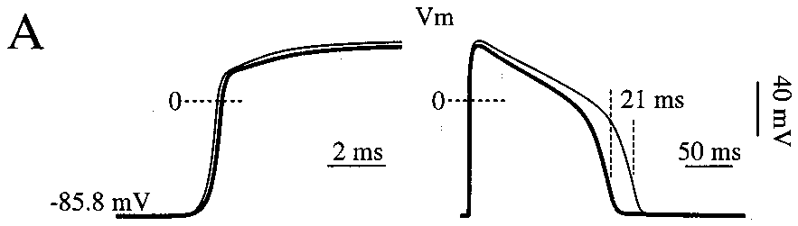 Figure 3: Mapping the h-gate spatial distribution and the identification of the reentrant "head" and "tail".
The Core Insight: Inactivation vs. Depolarization
The study found a fascinating duality in how Spontaneous Calcium Release (SCR) affects the heart:
- SCR above -40 mV (69% of cases): The calcium release causes rapid inactivation of L-type calcium currents (). This actually shortens the action potential duration (APD), leading to transient repolarization.
- SCR below -40 mV (31% of cases): The release triggers Delayed Afterdepolarizations (DADs). This prolongs the APD.
When these two effects happen in close proximity, they create a massive "dispersion of repolarization." Some cells recover early, others late. This dispersion is the "match" that lights the fire of wavefront fractionation—where one wave breaks into many, turning a stable tachycardia into chaotic fibrillation.
 Figure 5: Intracellular Calcium () and potential () waveforms across the functional center showing repeated SCR events.
Experiments: Testing Theoretical Thresholds
By adjusting the threshold for CICR () and calsequestrin buffer levels (), the authors showed they could modulate the frequency of these instabilities. Using an alternate formulation—the Jafri-Rice-Winslow (JRW) model—which handles subcellular subspace details more mechanistically, they confirmed that SCR remained a persistent destabilizer.
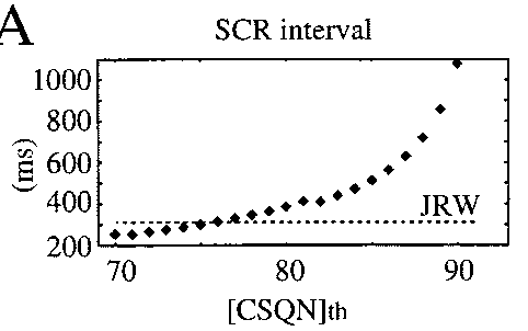 Figure 10: Comparative analysis of SCR intervals between LRd and JRW formulations under voltage clamp.
Conclusion: Beyond Electrical Mapping
The takeaway is profound: Functional reentry is not purely an electrical phenomenon. It is deeply coupled with the thermodynamics of calcium handling. The "leading circle" of the rotor inherently generates wave shapes that are optimized to trigger calcium instabilities.
Limitations & Future Work: The study used a monodomain representation and simplified geometry. Future research integrating 3D ventricular anatomy and discrete gap-junction modeling will be essential to see if these calcium "sparks" behave the same way in a heterogeneous organ.
Key Takeaway: Stabilizing the cell's calcium-handling proteins (like RyR or Calsequestrin) might be just as important as blocking sodium or potassium channels when treating high-rate arrhythmias.