
Deciphering the Aging Emotional Brain: A 3D Vector Field Tomography Power-Up for EEG
Age Effect in Human Brain Responses to Emotion Arousing Images: The EEG 3D-Vector Field Tomography Modeling Approach
This paper introduces a novel 3D Vector Field Tomography (3D-VFT) approach to model brain responses to emotional "anger" and "fear" stimuli using high-density 256-channel EEG. The study characterizes age-related differences in the N170 ERP component, identifying distinct neural activation patterns in young versus elderly populations.
Executive Summary
TL;DR: Researchers have developed a new way to "see" into the brain using High-Density EEG (256 channels) without needing an MRI for every subject. By applying 3D Vector Field Tomography (3D-VFT), they've mapped how young and elderly brains react to fear and anger. The verdict? The elderly react with much higher neural intensity (N170 amplitude) but show less "emotional differentiation" in their brain patterns compared to younger adults.
Academic Positioning: This work bridges the gap between high-temporal resolution EEG and high-spatial resolution fMRI. It introduces a physical-mathematical framework that treats the head as a bounded volume for field reconstruction, moving away from traditional, often biased, dipole fitting methods.
The "Inverse Problem" and Why Your Current Brain Maps Might Be Wrong
In neuroimaging, we face the "Inverse Problem": we measure electricity on the scalp, but we want to know what's happening inside.
- Prior Work Limitations: Most methods (like LORETA or dipole modeling) require "priors"—guesses about where the signal comes from or complex MRI scans of the subject's skull. If your model of the skull is slightly off, your brain map is way off.
- The Insight: The authors treat the brain as a vector field. By using the path-independency property of irrotational fields, they can reconstruct the internal state using only the boundary data (the EEG sensors).
Methodology: 3D Vector Field Tomography (3D-VFT)
The core of 3D-VFT is the discretization of the head into a grid of over 9,000 "cubic tiles." Instead of looking for a few "points" of electricity, the algorithm reconstructs the entire electrostatic field.
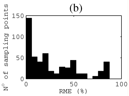 Figure 1: Comparison of 3D-VFT against Laplace’s PDE and ART. Note the significantly lower Mean Relative Magnitude Error (RME) and Angular Error (AE) for 3D-VFT.
The system solves a massive overdetermined linear system (mathbf{v} = \mathbf{K f}) where represents the potential differences between all possible sensor pairs. This provides a "regularization" effect that makes the reconstruction more stable than previous methods.
Key Findings: Age vs. Emotion
The study focused on the N170 component, a specific brain wave that occurs ~170ms after seeing a face.
1. The Intensity Gap
Elderly subjects showed a massive increase in N170 amplitude. While young adults showed ~-2.02μV for anger, the elderly spiked to -6.13μV. This suggests that as we age, the brain might recruit more neural resources to process negative emotional content—a phenomenon linked to the Socioemotional Selectivity Theory.
2. Spatial Mapping (The "Where")
Using 3D-VFT, the authors mapped the "hotspots" of the brain:
- Anger: Maximum activation in the Inferior Frontal Gyrus.
- Fear: Activation shifted toward the Superior Temporal Gyrus.
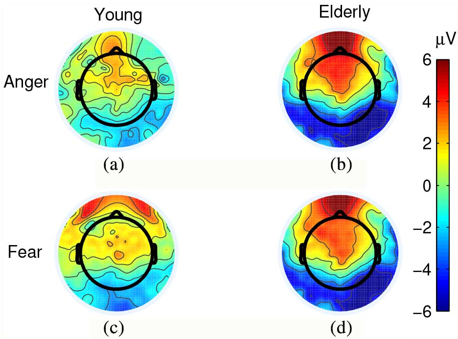 Figure 2: Coronal, Sagittal, and Axial slices showing the electrostatic field magnitude. Activation is clearly visible in the frontal and temporal lobes.
3. Emotional Homogeneity in the Elderly
Interestingly, while younger participants' brain maps looked very different depending on whether they saw fear or anger, the elderly participants' maps were much more similar. This suggests an age-related reduction in the ability to distinguish between different negative valences.
Critical Insight: Why This Matters for Clinical AI
This research isn't just about understanding emotions; it's about hardware-agnostic brain imaging. By proving that 3D-VFT can identify emotion-specific brain regions without subject-specific MRIs, it opens the door for:
- Affordable Clinical Monitoring: Tracking neurodegeneration (like Alzheimer’s) using just HD-EEG.
- Robust Emotional AI: Building systems that understand human affective states based on physical field modeling rather than "black-box" heuristics.
Conclusion
The N170 is more than a face-detection pulse; it is a window into how "emotional significance" is filtered by age. The 3D-VFT approach provides a mathematically rigorous way to peer through that window, showing that while our brain’s "functional atlas" for fear and anger stays largely the same as we age, the volume at which the brain "shouts" in response to these emotions increases dramatically.
Limitations: The study was conducted purely on female participants. Given known gender differences in emotional processing, expanding this to male cohorts is the next logical step for a universal brain model.