
Beyond Pixels: Bridging the Semantic Gap in Melanoma Detection with Ontology-Based Analysis
Automatic Skin Lesions Classification Using Ontology-Based Semantic Analysis of Optical Standard Images
This paper introduces an automated Computer-Aided Diagnosis (CAD) system for melanoma classification using an ontology-based semantic analysis of standard optical images. The method integrates a Bag-of-Words (BoW) model with Support Vector Machines (SVM) to bridge the gap between low-level image features and high-level medical concepts (ABCD rules), achieving SOTA sensitivity.
TL;DR
Diagnosis of melanoma via standard camera images is notoriously difficult due to poor contrast and lack of clinical context in raw data. This paper proposes a hybrid approach: using Ontologies to represent expert dermatological knowledge and SVM-driven Bag-of-Words to translate image features into meaningful clinical concepts. The result is a system that mimics a doctor's reasoning, achieving a remarkable 97.4% sensitivity.
Problem & Motivation: The "Black Box" vs. Clinical Reality
Most Computer-Aided Diagnosis (CAD) systems suffer from two main issues:
- The Hardware Gap: High-quality diagnosis usually requires a dermatoscope. Standard optical cameras (like those on smartphones) produce "noisy" data with lower saturation.
- The Interpretability Gap: Deep learning models often provide a diagnosis without explanation. In dermatology, the ABCD rule (Asymmetry, Border, Color, Differential structure) is the gold standard for human reasoning. Pure low-level feature extraction (texture/color) fails to capture these high-level clinical archetypes, leading to systems that doctors don't fully trust.
The authors' insight is simple but powerful: Don't just classify the image; model the doctor's language.
Methodology: Building a Clinical Ontology
The core of the system is an Ontology (O) defined by concepts (C) and semantic rules (R).
1. Feature Extraction to Semantic Annotation
Instead of feeding raw pixels to a classifier, the authors extract specific geometric and colorimetric features:
- Asymmetry (CA): Combined Central/Axial shape asymmetry and Color asymmetry using Chi-square distance.
- Border (CB): Measuring "Compactness" and "Shape Signatures" to quantify irregularities.
- Color (CC): Using K-means to determine the number of distinct color clusters.
- Differential Structures (CD): Utilizing GLCM (Gray Level Co-occurrence Matrix) for texture analysis.
2. The Bag-of-Words (BoW) Bridge
These features are processed by an SVM to assign "words" to the lesion—for example, a lesion might be labeled as "Highly Asymmetric" or having "Coarse Irregularities."
 Figure 1: The workflow from raw image to semantic rule application.
3. Rule-Based Decision Making
The final decision is guided by the ABCD Score. The system calculates a Total Score (): If , the lesion is flagged as Melanoma. This isn't just a statistical guess; it's a derivation based on modeled clinical criteria.
Experiments & Results: Sensitivity is King
The model was evaluated on 206 images from DermQuest and DermIS. In the world of cancer, Sensitivity (not missing a case) is more important than specificity (avoiding false alarms).
| Method | Sensitivity | Accuracy |
|---|---|---|
| Previous works (Low-level) | 61% - 92% | 71% - 92% |
| This Work (Ontology-Based) | 97.4% | 76.9% |
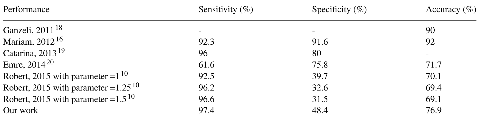 Table 1: Comparative study showing the superior sensitivity of the proposed approach.
While the accuracy (76.9%) is lower than some texture-only models, the 97.4% Sensitivity indicates that this system is much safer for a preliminary screening tool. It rarely misses a malignant lesion, which is the primary goal of any medical CAD system.
Critical Analysis & Conclusion
Takeaway
The paper proves that a Knowledge-Based System can significantly improve the safety profile (sensitivity) of medical AI. By forcing the AI to "think" in terms of the ABCD rule, the authors created a bridge between machine learning and human expertise.
Limitations & Future Work
The main drawback is Specificity (48.4%). The current rules are "too permissive," leading to many false positives (benign lesions flagged as malignant). The authors suggest moving toward Fuzzy Logic in future iterations to handle the "gray areas" between categories (e.g., the transition from "Medium" to "High" asymmetry) more gracefully.
Final Thought
As we move deeper into the era of Black-Box Deep Learning, this paper serves as a reminder that domain-specific structures (like Ontologies) are still vital for creating systems that are not just accurate, but clinically responsible.