
Deciphering Vascular Aging: The Strong Link Between CIMT and Diastolic Augmentation Index
Relation between Carotid Intima-Media Thickness and diastolic augmentation index in Chinese population
This clinical study investigates the relationship between subclinical atherosclerosis, measured via Carotid Intima-Media Thickness (CIMT), and arterial stiffness, quantified by the Radial Diastolic Augmentation Index (DAI), in a Chinese outpatient population. Using tonometric and ultrasound methods, the study establishes DAI as a potent predictor of vascular health alongside traditional markers like PWV and AI.
TL;DR
This study investigates the interplay between the "structure" and "function" of the arterial system in a Chinese cohort. By comparing Carotid Intima-Media Thickness (CIMT)—a mirror of atherosclerosis—with the Diastolic Augmentation Index (DAI)—a measure of arterial stiffness—the researchers found that DAI is a highly significant, independent predictor of vascular damage, potentially outperforming traditional metrics in specific clinical contexts.
Problem & Motivation: Why Look Beyond Pulse Wave Velocity?
In the world of cardiovascular diagnostics, we often separate structural damage (like plaque buildup and wall thickening) from functional stiffening (how hard the "pipes" have become).
- CIMT is the gold standard for structural imaging via ultrasound.
- PWV (Pulse Wave Velocity) and AI (Augmentation Index) are the traditional functional metrics.
However, PWV can be technically difficult to measure accurately in older or obese populations due to the difficulty of capturing femoral pulses. The authors focus on DAI, derived from simple radial artery tonometry (the pulse at your wrist), to see if it can serve as a robust surrogate for the structural decay seen in carotid arteries.
Methodology: Bridging Ultrasound and Tonometry
The study recruited 65 participants (mean age 51) and employed a dual-diagnostic approach:
- Structural Assessment: Automatic CIMT measurement using a 5-10 MHz linear transducer.
- Functional Assessment: Radial pulse wave analysis using the IIM-2010A device.
As shown in the waveform analysis, the team focused on the diastolic portion of the pulse wave. While the standard Augmentation Index (AI) focuses on the systolic peak (P2), the DAI focuses on the late-systolic/early-diastolic peak (P3), providing a unique window into how pressure waves reflect back toward the heart.
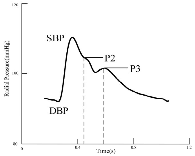 Figure 1: Definition of SBP, DBP, P2 (Systolic Peak), and P3 (used for DAI).
Key Results: A Robust Correlation
The results revealed a striking "inverse mirror" relationship. As the carotid artery walls thicken (higher CIMT), the diastolic augmentation index (DAI) drops significantly.
- The Quantitative Evidence: The correlation coefficient between CIMT and DAI (r = -0.636) was notably stronger than that of CIMT and PWV (r = 0.396).
- Predictive Power: In a stepwise regression, DAI remained a significant predictor of CIMT even after adjusting for age and blood pressure.
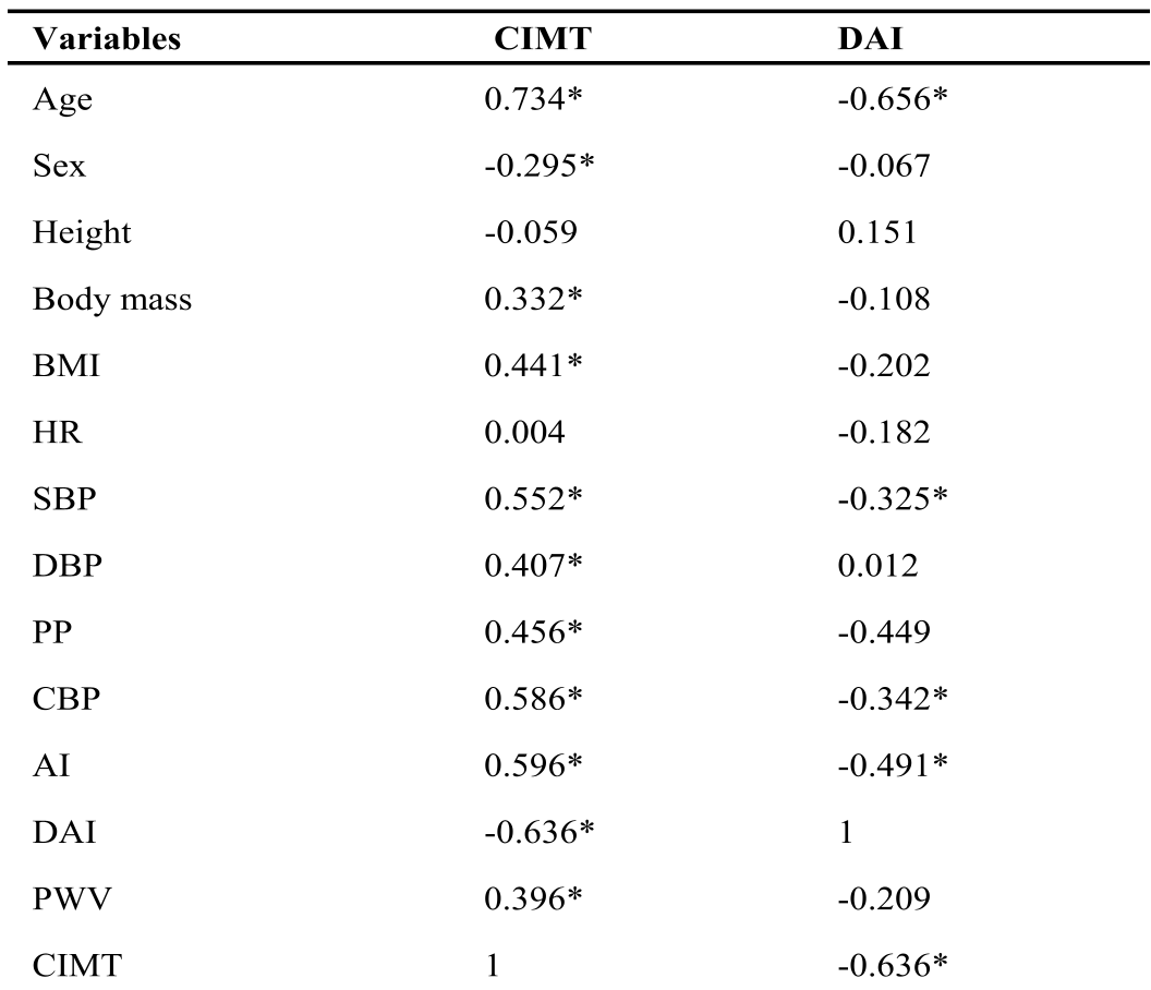 Table 1: Univariate correlations showing DAI as a top associate of CIMT.
Critical Analysis: Why DAI Matters
The study suggests that DAI is more sensitive to the structural changes of atherosclerosis than PWV in older Chinese populations. The authors' insight is rooted in hemodynamics: as arteries stiffen, the reflected pressure wave returns earlier. While AI measures this in systole, DAI captures the tail end of this reflection.
The fact that DAI decreases with age while AI increases highlights a "crossover" in how we should measure vascular aging across different life stages. In older subjects, where CIMT is already elevated, DAI provides a clearer resolution of the arterial tree's health.
Limitations
- Sample Size: With 65 subjects, the study is a pilot-scale investigation.
- Gender Bias: The cohort had significantly more females than males, and the researchers noted that estrogen deficiency (menopause) could influence arterial stiffness independently.
Future Outlook
This research elevates DAI from a "novel experimental parameter" to a "clinically relevant marker." Future diagnostic tools for primary care might favor radial tonometry—which is as simple as wearing a sophisticated watch—over carotid ultrasounds to identify patients at high risk for stroke or myocardial infarction before visible plaques even form.
Takeaway: If you want to know the state of a patient's carotid "structure," looking at their radial "function" via DAI is a highly reliable and efficient shortcut.