
COMPARE: Bridging the Gap Between Voxel-Wise Noise and Anatomical Rigidity in Brain Classification
COMPARE: Classification of Morphological Patterns Using Adaptive Regional Elements
The paper introduces COMPARE (Classification of Morphological Patterns Using Adaptive Regional Elements), a novel framework for neuroimaging classification. It combines mass-preserving high-dimensional image warping with a watershed-based adaptive regional feature extraction and SVM-based selection to achieve high-accuracy identification of schizophrenia from structural MR images.
TL;DR
The COMPARE framework revolutionizes structural MRI analysis by moving away from "fixed" anatomical regions. Instead, it uses a watershed-based adaptive grouping to find "morphological signatures" unique to a disease. By combining mass-preserving transformations with Support Vector Machines (SVM), it achieved over 90% accuracy in detecting schizophrenia, outperforming traditional wavelet and ROI methods.
Context: The Multivariate Challenge in Neuroimaging
In the world of computational anatomy, researchers often face a dilemma. You can look at individual pixels (voxels), which is precise but extremely noisy, or you can look at large predefined Regions of Interest (ROIs), which are robust but might miss subtle, "oddly shaped" abnormalities that don't follow anatomical borders.
The authors of this paper argue that schizophrenia doesn't care about your atlas boundaries. To find it, we need a method that is both multivariate (looking at relationships between regions) and adaptive (letting the data define the regions).
Methodology: How COMPARE Works
The "magic" of COMPARE lies in its three-stage pipeline:
1. Mass-Preserving Warping (RAVENS)
Before comparing brains, they must be aligned. The authors use the HAMMER algorithm to warp individual brains to a template. Crucially, they use a "mass-preserving" framework. If a region is compressed to fit the template, its "tissue density" increases—ensuring the total volume of gray matter or white matter remains constant for measurement.
2. Adaptive Regional Grouping (The Watershed approach)
Instead of using a standard atlas, COMPARE calculates a Discrimination and Robustness Measure (DRM) for every voxel.
- Relevance: How well does this voxel correlate with the disease?
- Robustness: Is the measure consistent across neighbors (Spatial Consistency)?
They then apply a Watershed Segmentation to this DRM map. This groups voxels into irregularly shaped "functional" regions that are most likely to show disease-related changes.
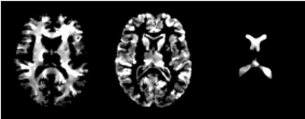 Figure 1: Visual representation of tissue density maps and the adaptive partitioning.
3. Feature Selection & SVM
Since even these regions can be redundant, the authors use SVM-RFE (Recursive Feature Elimination). This prunes the features down to a "morphological signature"—a small set of measurements that provide the maximum separation between "Healthy" and "Schizophrenia" in a high-dimensional space.
Experimental Results: SOTA Performance
The researchers tested COMPARE on two datasets (Male and Female). The results were strikingly stable compared to other techniques:
| Method | Female (Accuracy) | Male (Accuracy) |
|---|---|---|
| COMPARE (Proposed) | 91.8% | 90.8% |
| Wavelet-based | 86.9% | 85.1% |
| ROI-based | 80.3% | 80.5% |
| PCA | 82.0% | 73.6% |
The stability of the SVM performance across different kernel sizes (as seen in Figure 4) proves that the extracted features are inherently discriminative and not just a result of hyperparameter tuning.
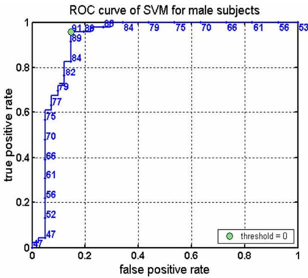 Figure 2: Accuracy plots showing that SVM-based selection (right) provides much more stable results than simple ranking (left).
Deep Insight: Why Does it Work?
The core reason COMPARE wins is its Spatial Adaptivity. Traditional Gaussian smoothing (used in Voxel-Based Morphometry) blurs the brain uniformly. COMPARE, however, uses its watershed approach to "cluster" voxels only where the signal is strong and robust. This essentially acts as a "statistical edge detector," identifying regions like the Hippocampi as key discriminators, consistent with long-standing clinical findings in schizophrenia.
Conclusion & Future Outlook
COMPARE demonstrates that in medical imaging, "where" you look is just as important as "how" you look. By allowing the pathology to define the regions of interest, the model achieves near-diagnostic levels of accuracy.
Limitations: The study relies on a relatively small sample size (typical for its era), and the "leave-one-out" cross-validation can sometimes be optimistic.
Future Work: Integrating this method with modern Deep Learning (like 3D CNNs or Transformers) could allow for even more complex, non-linear feature extraction that doesn't rely on manual watershed segmentation.