
[arXiv 2022] DDM: Generative 4D Cardiac MRI via Diffusion-Guided Deformation
Diffusion Deformable Model for 4D Temporal Medical Image Generation
The paper introduces the Diffusion Deformable Model (DDM), a novel 4D medical image generation framework that combines Denoising Diffusion Probabilistic Models (DDPM) with deformable registration. It achieves State-of-the-Art performance in generating intermediate 3D cardiac volumes between diastolic and systolic phases by interpolating a learned latent code.
Executive Summary
TL;DR: The Diffusion Deformable Model (DDM) tackles the challenge of generating high-fidelity intermediate 3D volumes in temporal (4D) medical sequences. By utilizing a diffusion-based latent code to guide a deformation engine, it generates continuous, topology-preserving trajectories between two anatomical states (e.g., end-diastole to end-systole).
Context: Within the medical imaging landscape, this work marks a shift from purely discriminative registration (finding a map) or standard generative modeling (creating pixels from scratch) toward a hybrid approach: Generative Deformation.
Problem & Motivation: The Limits of Linear Motion
Capturing 4D dynamics (3D + time) in MRI is time-intensive and clinically taxing. While researchers have tried GANs to "hallucinate" missing frames, GANs often fail clinical "sanity checks" by introducing artificial structures.
Registration-based methods like VoxelMorph (VM) are safer because they warp existing anatomy (preserving topology), but they have a fatal flaw: to generate intermediate frames, they usually just scale the final displacement field (). This assumes every part of the heart moves in a straight line at a constant speed, which is physiologically incorrect.
Methodology: Diffusion meets Deformation
The authors propose a two-stage end-to-end pipeline:
- Diffusion Module: Instead of generating an image, this module learns to estimate a "score" or a latent code . This code encapsulates the high-dimensional spatial "force" required to transition from Source to Target .
- Deformation Module: This module takes the source image and the latent code . It uses the code to produce a deformation field . By scaling the latent code (), the model allows for smooth, non-linear interpolation.
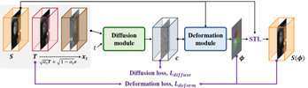
The core insight is that the diffusion process naturally learns the gradients (scores) of the data distribution. By conditioning this on the source/target pair, the latent code acts as a "velocity" guide that is more expressive than simple linear displacement.
Experiments & Results
The model was validated on the ACDC dataset (4D Cardiac MRI).
Quantitative SOTA Performance
DDM consistently outperformed standard VoxelMorph (VM) and its diffeomorphic variant (VM-diff):
- PSNR: 30.725 dB (Highest among baselines)
- Dice Score: 0.802 (Superior anatomical alignment)
- Inference Speed: ~0.45s per volume, making it 6x faster than VM-diff while maintaining better performance.
Visual Fidelity
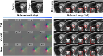
As shown in the comparison below, while VM produces "mechanical-looking" linear movements, DDM captures the complex thickening and contraction of the myocardium more naturally.
Critical Analysis & Conclusion
Takeaway
The DDM framework proves that we don't need to choose between the "realism" of Diffusion models and the "structural integrity" of Registration. By using Diffusion to parameterize the deformation rather than the pixels, we get the best of both worlds: realistic temporal dynamics and guaranteed anatomical topology.
Limitations & Future Work
- Boundary Conditions: While the geodesic path is plausible, the model doesn't explicitly enforce physical constraints (like incompressible fluid dynamics of heart tissue).
- Extension: The current work focuses on two-endpoint interpolation (). Future work could extend this to multi-point trajectories for full cycle (diastole-systole-diastole) modeling.
DDM stands as a significant step toward "Virtual 4D Imaging," where short scans can be computationally expanded into high-resolution physiological movies.