
Extreme Learning Machine: Redefining Speed and Accuracy in Fatty Liver Stratification
Extreme Learning Machine Framework for Risk Stratification of Fatty Liver Disease Using Ultrasound Tissue Characterization
This study presents a reliable and high-speed risk stratification system for Fatty Liver Disease (FLD) titled "Symtosis," utilizing an Extreme Learning Machine (ELM) framework applied to ultrasound (US) tissue characterization. By training a Single Layer Feed-Forward Neural Network (SLFFNN) on 46 grayscale features, the method achieves a state-of-the-art accuracy of 96.75% and an AUC of 0.97 on sub-sampled datasets.
TL;DR
Fatty Liver Disease (FLD) is a global health challenge requiring non-invasive, fast, and accurate diagnostic tools. This paper introduces the Symtosis system, which replaces traditional, slow iterative classifiers like SVM with an Extreme Learning Machine (ELM). The results are striking: a 96.75% accuracy and a 40% speed-up over conventional methods, all while maintaining near-perfect reliability.
The Bottleneck in Clinical CADx
Computer-Aided Diagnosis (CADx) systems for ultrasound imaging have historically relied on kernels (SVM) or backpropagation (BPNN). While effective, these methods are "mathematically stressed." SVMs require intensive computation to find support vectors in high-dimensional spaces, and BPNNs require thousands of iterations to converge. In a clinical setting, where efficiency is as vital as accuracy, these bottlenecks hinder real-time risk stratification.
Why ELM Works: The Power of Randomness and Pseudo-Inverses
The core intuition behind the ELM-based Symtosis system is the decoupling of weight layers. Unlike standard Neural Networks, ELM does not tune the input-to-hidden layer weights; instead, it randomly initializes them.
The Single-Pass Logic
The only weights that are learned are those between the hidden and output layers. This is achieved through a "Single-Pass" approach:
- Feature Mapping: 46 grayscale features (Gabor, GLCM, GRLM) are mapped into a hidden layer.
- Least-Squares Solution: Instead of gradient descent, the system uses the Moore-Penrose generalized inverse to find the global optimum in one mathematical step.
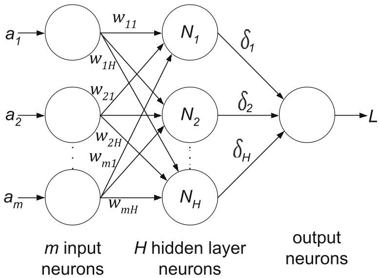 Fig 1: The SLFFNN architecture of the ELM paradigm within the Symtosis framework.
Experimental Mastery: S0, S4, and S8
To overcome the limitation of a small clinical dataset (63 patients), the authors employed a sub-sampling strategy:
- S0: The original images.
- S4: Images split into 4 parts.
- S8: Images split into 64 parts (yielding 4,032 samples).
This allowed the ELM to demonstrate its superior generalization capabilities as the data size increased, a common requirement for modern clinical validation.
Performance Benchmarking
The ELM was pitted against SVM across four Cross-Validation (CV) protocols. The superiority was consistent:
- Accuracy: ELM hit 96.75% (K10, S8) vs. SVM’s 89.01%.
- Area Under Curve (AUC): ELM reached 0.97, indicating nearly perfect classification.
- Speed: Training and testing time for ELM were consistently lower, providing a 40% average speed-up.
 Table 1: Quantifying the transition from SVM to ELM—Higher accuracy and sensitivity across all protocols.
Critical Insight: ELM vs. BPNN
One might argue that complex Back Propagation Neural Networks (BPNN) could achieve similar or slightly higher accuracy (97.6%). However, the authors emphasize that ELM achieves its 96.7% using one-third of the features and one-tenth of the layer complexity of a BPNN. This "Efficiency-Performance" trade-off is the true value proposition of the ELM framework.
Conclusion & Future Outlook
The Symtosis study proves that ELM is not just a theoretical curiosity but a robust tool for medical tissue characterization. By achieving high reliability (99%) and stability (SD < 5%), it offers a path forward for real-time diagnostic hardware.
Future Directions: The next frontier for this framework is its application in "Big Data" medical contexts and the integration of deep learning feature extractors (like CNNs) to replace manual grayscale feature engineering.