
Automated Oocyte Grading: Bridging the Gap Between Morphology and Machine Learning
9271_Grading of mammalian cumulus oocyte complexes using machine learning for in vitro embryo culture.
The paper presents a semi-automatic machine learning framework for grading mammalian Cumulus Oocyte Complexes (COCs) to improve in vitro fertilization (IVF). It combines multi-object parametric segmentation (Active Contours and Region Growing) with a Random Forest classifier to categorize oocytes into four quality grades (A-D).
TL;DR
Precise oocyte selection is the cornerstone of successful in vitro fertilization (IVF), yet it remains a subjective manual task. This research introduces a semi-automated pipeline that uses Active Contours and Random Forests to grade mammalian Cumulus Oocyte Complexes (COCs) with 88.35% accuracy, demonstrating that mathematical "contour-based" features far outperform traditional textural analysis in predicting biological competence.
The Subjectivity Crisis in Embryology
In the world of IVF, the "quality" of an oocyte is often determined by the thickness of its cumulus cell layers and the homogeneity of its ooplasm. Currently, this is a visual call made by biologists under a microscope. The problem? One expert’s "Grade B" might be another’s "Grade C." This subjectivity leads to batch-to-batch variability and inconsistent embryo production. While molecular assays (like gene expression profiling) are precise, they are essentially "destructive" or too slow for real-time selection. There is an urgent need for a non-invasive, standardized "digital eye."
Methodology: Seeing Beyond Pixels
The authors recognized that the most critical indicators of oocyte health lie in the spatial relationship between the nucleus and its surrounding cumulus "investment." Their approach follows a rigorous three-step technical pipeline:
1. Multi-Object Segmentation
Instead of treating the oocyte as a single blob, the framework segments two concentric regions:
- The Outer Boundary: Captured using Active Contours (Snakes), which iteratively fit a curve to the outer edge of the cumulus layer.
- The Nucleus: Isolated via a Region Growing (RG) algorithm, initialized by the geometric center of the outer boundary.
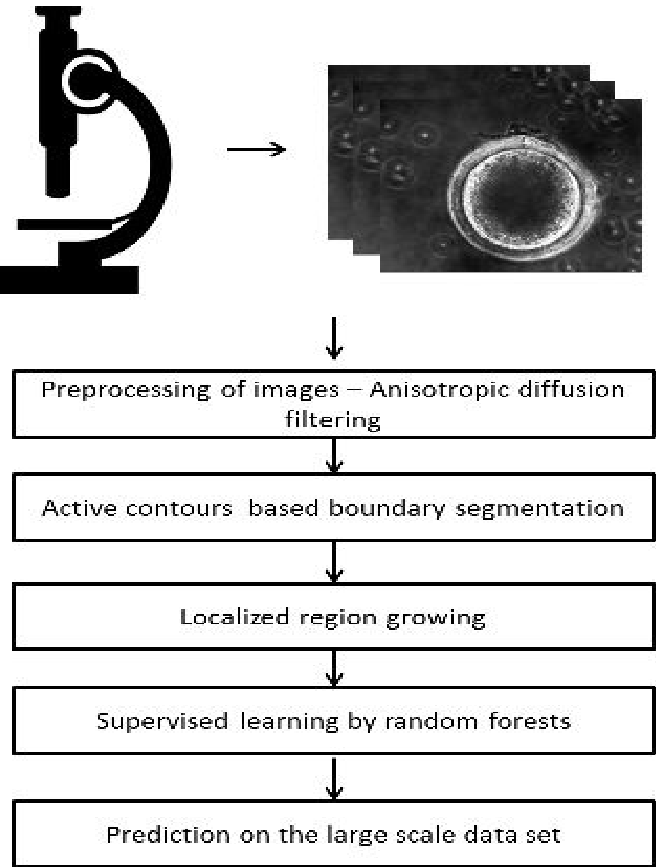 Figure 1: The proposed workflow, from image acquisition to the Random Forest ensemble.
2. Feature Engineering: The "Secret Sauce"
The study compared two types of features:
- Texture-based: Local Binary Patterns (LBP) and Gradients (the "feel" of the image).
- Contour-based: The radius ratio between the inner and outer circles and the relative area of the cumulus layer.
3. Classification via Random Forest
The researchers chose Random Forest (RF) over SVMs or Neural Networks due to its inherent robustness to small, unbalanced datasets—a common reality in specialized biological imaging.
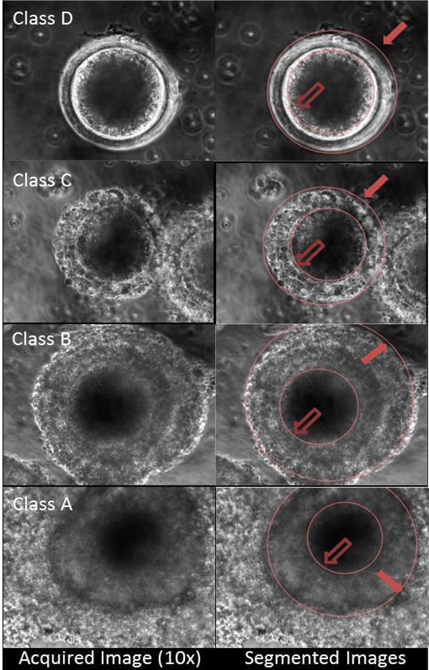 Figure 2: Visualization of parametric curves fitting the nucleus (inner) and cellular boundary (outer) across different grades.
Experimental Performance
The model was tested on 80 mammalian oocytes with a 30/70 train-test split. The findings were revealing:
- Overall Accuracy: 88.35%.
- The Power of Geometry: Using only texture features yielded only 75% accuracy. Adding the contour-based features (derived from the segmentation step) pushed the performance by over 13%.
- Grade Extremes: The system was perfect (100% accuracy) at identifying Grade A (highest quality) and Grade D (lowest quality) oocytes. The minor errors occurred only in the subtle transition between Grades B and C.
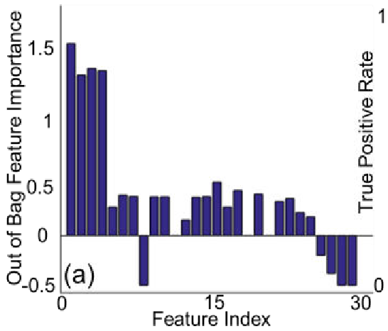 Figure 4: Analysis of feature importance (a) showing that segmentation-derived features were the most predictive.
Academic Insight: Why it Works
The high performance of this framework stems from its Inductive Bias. By forcing the model to look at the ratio of the nucleus to the cumulus layer (a biologically significant metric), the authors provided the machine learning model with a "structural hint" that raw pixel data alone couldn't easily learn. This proves that in medical imaging, "Feature Engineering" informed by domain expertise is still a potent weapon, especially when data is scarce.
Conclusion & Future Outlook
This work successfully transforms a subjective biological assessment into a quantitative engineering protocol. While the current framework is "static" (analyzing still images), the authors suggest that moving toward dynamic evaluation (video tracking) could further refine accuracy. For the IVF industry, this translates to a scalable, standardized tool that reduces the "human factor," ultimately leading to higher success rates in embryo culture.
Limitations: The dataset size (80 samples) is small by modern deep learning standards. Moving forward, validating this on larger, multi-species datasets will be essential for clinical adoption.