
HFμDI: Breaking the Transparency Barrier in Adult Zebrafish Vascular Imaging
165_In Vivo Visualization of Vasculature in Adult Zebr
This paper introduces High-Frequency Micro-Doppler Imaging (HFμDI), an in vivo vascular mapping technique for adult zebrafish using a 40-MHz ultrafast ultrasound system. By combining high-frequency transducers with adaptive SVD-based clutter filtering, the method achieves label-free visualization of small peripheral vessels with a resolution of 36 μm and a penetration depth of 4 mm.
TL;DR
Researchers have developed a breakthrough imaging technique called High-Frequency Micro-Doppler Imaging (HFμDI). By pushing ultrasound frequencies to 40 MHz and utilizing ultrafast plane-wave transmissions, they can now map the entire 3D vascular system of an adult zebrafish in vivo. Unlike optical methods, it doesn't require transparent "albino" mutants or fluorescent labels, achieving a 36 μm resolution and 4 mm depth entirely label-free.
Context: Why the Adult Zebrafish is an Imaging Nightmare
Zebrafish are the "rockstars" of developmental biology and cancer research. However, they have a major limitation: as they mature, they develop scales and stripes that make them opaque.
- Optical limitations: Specialized microscopes (Confocal, OCT) work great for transparent larvae but can't see through the dense tissue of an 8-month-old adult.
- Traditional Ultrasound limitations: Standard Doppler ultrasound lacks the sensitivity to catch the slow, minuscule blood flow in peripheral vessels (3-4 mm/s), often drowning these signals in "clutter" (tissue movement).
The Core Innovation: High Frequency meets Ultrafast SVD
The researchers solved this by combining two cutting-edge technologies:
- 40-MHz Ultrafast Imaging: Using the highest frequency currently available for ultrafast systems, they achieved a high frame rate (500 Hz) that captures thousands of snapshots of red blood cell movements.
- Adaptive Spatiotemporal SVD Filtering: This is the mathematical "magic" that separates blood from bone. By treating the stack of images as a large matrix, they apply Singular Value Decomposition (SVD) to filter out static or slow-moving tissue (high energy) and electronic noise (random frequency), leaving only the distinct signature of flowing blood.
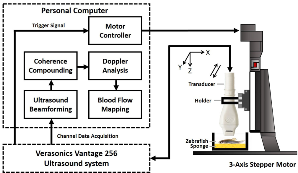 Figure 1: The schematic shows the 40-MHz array transducer setup integrated with a 3-axis motor for 3D reconstruction.
Methodology: Tuning the "Microscope"
The study meticulously optimized two critical parameters:
- Tilted Plane Wave Angles: They found that 7 angles provided the best balance between Image SNR (Signal-to-Noise Ratio) and acquisition speed.
- SVD Block Size: An adaptive block size of 64 pixels was found to be the "sweet spot" for distinguishing peripheral vessels without blurring the signal.
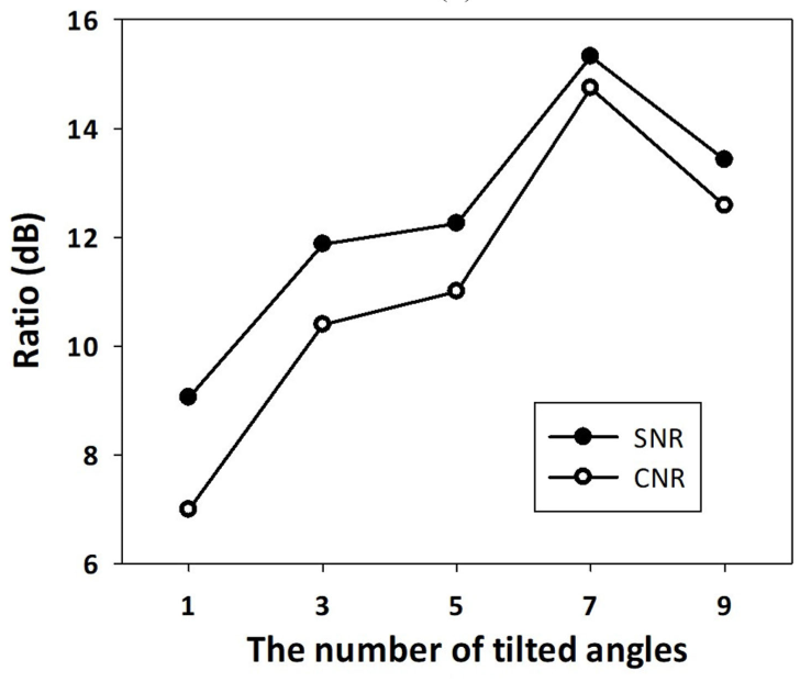 Figure 3: The thresholding strategy uses both the magnitude of singular values and Doppler frequency to isolate blood signals.
Experimental Validation: Seeing the Unseen
The system was tested on 8-month-old wild-type zebrafish. The results were stunning:
- 3D Visualization: For the first time, researchers could see the Intersegmental Vessels (Se), Dorsal Aorta (DA), and the complex network connecting them in a fully grown, opaque fish.
- Physiological Sensitivity: To prove sensitivity, they changed the water temperature. HFμDI was precise enough to detect a 14.4% decrease in vessel diameter as the fish underwent vasoconstriction at 20°C compared to 30°C.
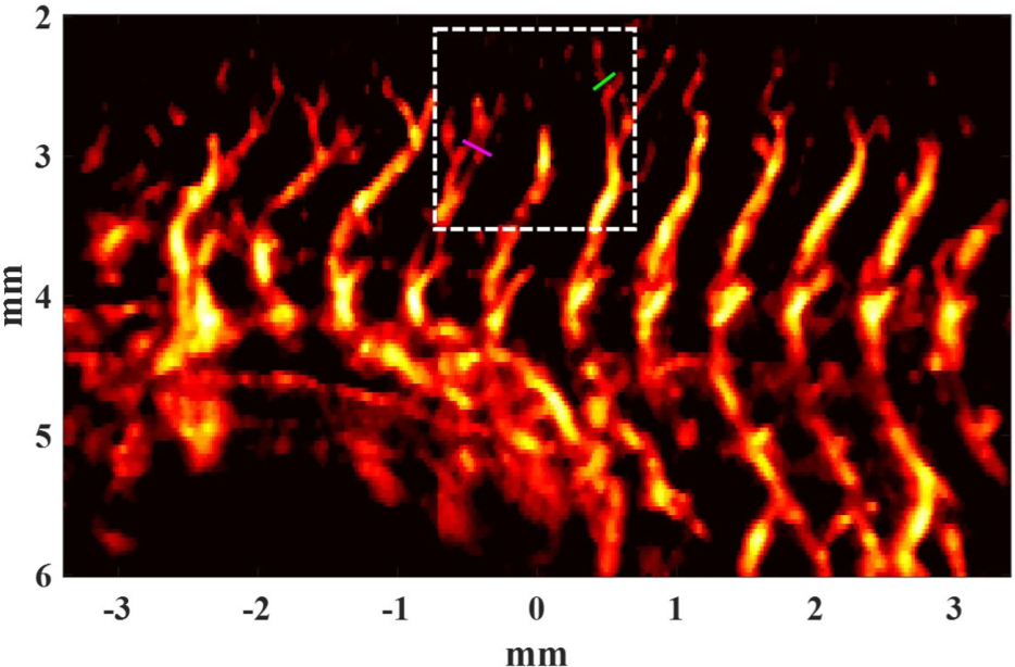 Figure 6: (b) Power Doppler images showing the intricate Se, PAV, and DLAV vessels. (d) Color Doppler indicating flow direction—red for arteries and blue for veins.
Deep Insight: A New Standard for Microcirculation
The significance of HFμDI lies in its independence. Most high-resolution zebrafish studies rely on transgenic "reporter" lines that express green fluorescent protein (eGFP). HFμDI works on any fish because it relies on the physical backscattering of red blood cells (RBCs). Furthermore, at 40 MHz, the backscattering of RBCs is significantly stronger due to Rayleigh scattering (which scales with the 4th power of frequency), allowing for vessel detection down to 36 μm without needing to inject microbubbles.
Conclusion & Future Outlook
Takeaway: HFμDI provides a unique "macro-to-micro" perspective. It matches the resolution of complex optical systems (like OR-PAM) while offering the depth and velocity-sensing capabilities of ultrasound.
Limitations: While 36 μm is impressive, it is still above the single-capillary level (typical zebrafish capillaries are <10 μm). Future work combining this high-frequency approach with Super-Resolution Localization Microscopy could potentially reach the 5-10 μm range.
Prognosis: This technology isn't just for fish. The authors hint at clinical applications in human microcirculation, such as monitoring tumor angiogenesis in skin cancer or evaluating wound healing in superficial tissues.