
Deciphering the Powerhouse: Advanced Protocols for Plant Mitochondrial Fractionation and Proteomics
Isolation and Subfractionation of Mitochondria from Plants
This paper provides a comprehensive methodology for the isolation, purification, and subfractionation of plant mitochondria, specifically detailing techniques like Percoll/sucrose density gradient centrifugation and Blue-Native PAGE. It establishes a standardized framework for proteome analysis and "in organello" translation, achieving high-purity organelle preparations suitable for mass spectrometry.
TL;DR
Plant mitochondria are metabolic hubs that differ significantly from their mammalian counterparts, containing unique enzymes and rigid structural hurdles. This seminal methodology paper by Millar et al. provides a masterclass in isolating, purifying, and subfractionating these organelles. By combining density gradient centrifugation with Blue-Native PAGE and Mass Spectrometry, the authors move beyond simple "crude extracts" to high-resolution proteomic mapping of the mitochondrial compartments.
The Plant Problem: Beyond the Cell Wall
Why can't we just use animal mitochondrial protocols for plants? The paper identifies three major roadblocks:
- Mechanical Armor: The plant cell wall requires aggressive homogenization (mortar/pestle or blenders), which risks rupturing the delicate mitochondrial membranes.
- Chemical Interference: Rupturing vacuoles releases acids and phenolics that can instantly "tan" or deactivate mitochondrial proteins.
- The Green Contaminant: In leaf tissues, chloroplasts and thylakoids share similar densities with mitochondria, making clean separation nearly impossible without specialized gradients.
Methodology: The Art of Gradient Purification
The core of the paper revolves around two primary purification routes: Sucrose and Percoll gradients.
1. Density Gradient Strategy
The authors favor Percoll (colloidal silica) for rapid purification. Unlike sucrose, Percoll allows for iso-osmotic conditions, preventing the "osmotic shock" that causes mitochondria to swell and burst. For green tissues, they suggest a 0–5% PVP-40 gradient within the Percoll to specifically scrub thylakoid contamination.
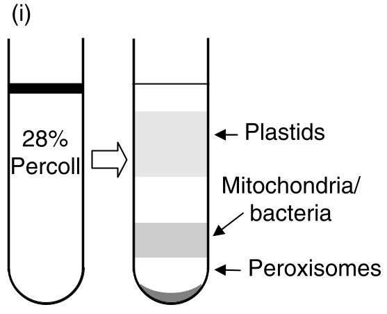 Fig. 1: Isolation profiles showing the separation of mitochondria from plastids and peroxisomes.
2. Subfractionation: Mapping the Four Compartments
To understand protein localization, the paper details how to "peel" the organelle into:
- Outer Membrane (OM)
- Inner Membrane (IM)
- Matrix (MA)
- Intermembrane Space (IMS)
This is achieved through carefully timed osmotic shocks using 10-mM or 86-mM sucrose, selectively rupturing the outer membrane while leaving "mitoplasts" (IM + MA) intact for further separation.
Analyzing the Proteome: Blue-Native PAGE
One of the most powerful techniques discussed is Blue Native Polyacrylamide Gel Electrophoresis (BN-PAGE). Unlike standard SDS-PAGE which denatures proteins, BN-PAGE uses Coomassie Blue G-250 to confer a negative charge to intact protein complexes.
This allows researchers to see the "Electron Transport Chain (ETC)" in its native, functional state. The authors used this to identify plant-specific subunits in Complex I (NADH-Q oxidoreductase) and Complex II (Succinate dehydrogenase) that simply do not exist in other kingdoms.
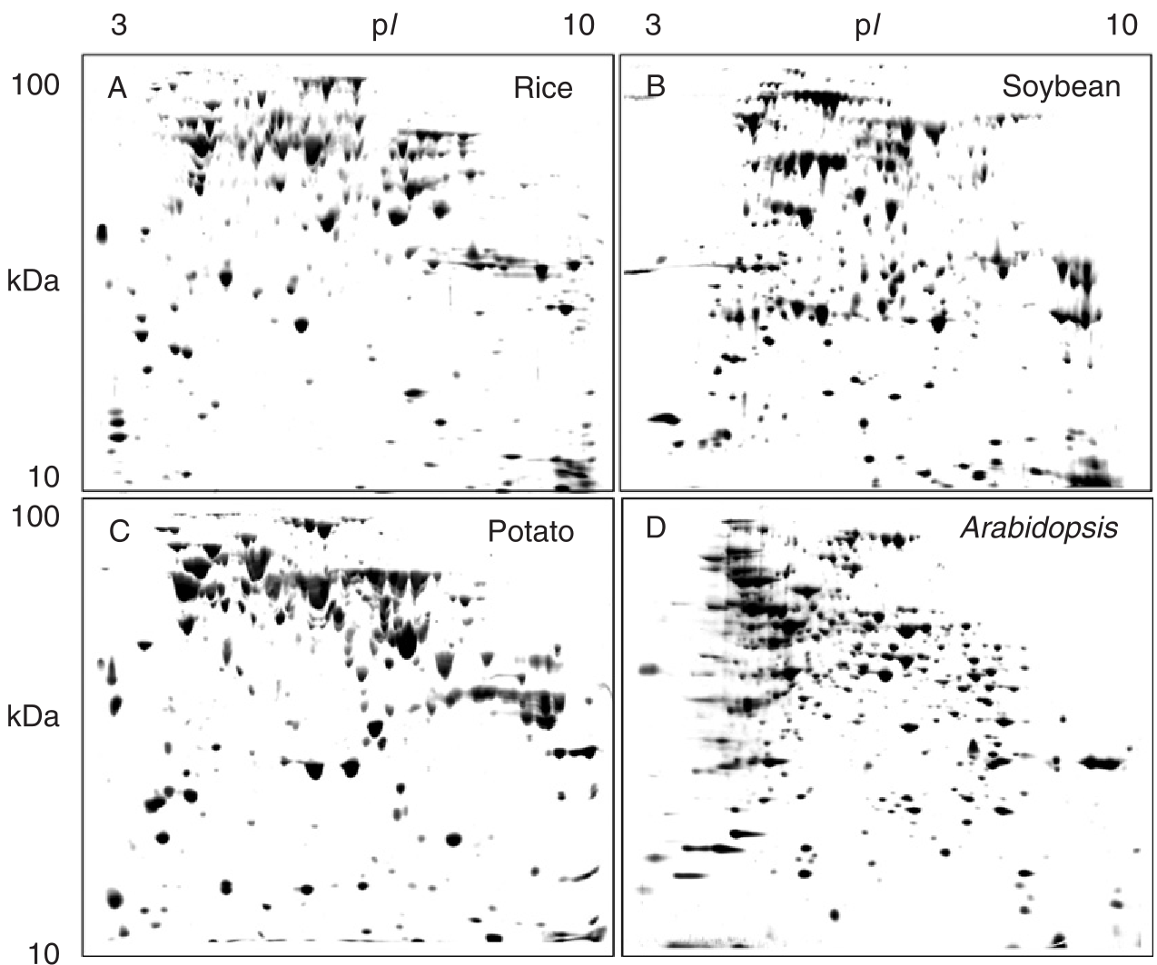 Fig. 6: 2D analysis (BN-PAGE followed by SDS-PAGE) revealing the subunit structure of intact respiratory complexes.
Experiments & Quantitative Purity
The paper sets high standards for "Integrity Determinations." They suggest two critical checks:
- Cytochrome c Latency: Since the inner membrane is impermeable to Cytochrome c, measuring oxidase activity before and after detergent addition allows for a precise "integrity percentage" (typically >90%).
- Marker Enzyme Assays: Using Fumarase (Matrix) vs. Catalase (Peroxisomal contamination) to validate that the result is truly mitochondrial.
Critical Analysis & Future Outlook
The value of this paper lies in its scalability. Whether you are working with 10kg of potato tubers for protein purification or 3g of transgenic Arabidopsis tissue for screening, the authors provide a modified "Mini-Prep" protocol.
Limitations: While the paper is a gold standard for biochemistry, it acknowledges that "gel-free" proteomics (purely LC-MS based) is the next frontier for identifying low-abundance signaling proteins that 2D gels often miss.
Conclusion: For any researcher looking to understand how plants generate energy under stress or how the mitochondrial genome communicates with the nucleus, these isolation protocols are the essential prerequisite. They transform the organelle from a "black box" into a series of clearly defined, assayable protein modules.