
RadiSim-CL: Training AI to "Think" Like a Professional Radiologist
Learning like a radiologist: a medical vision-language model for radiological image analysis via curriculum learning
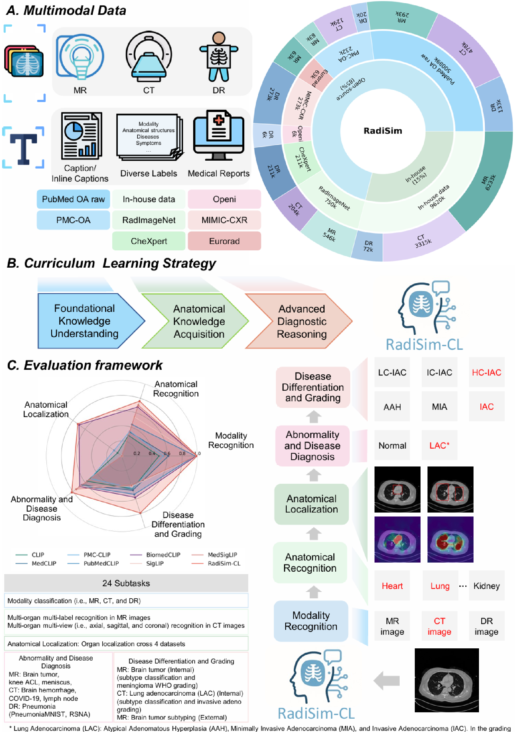
The paper introduces RadiSim-CL, a specialized Medical Vision-Language Model (MVLM) designed to mimic the three-phase learning trajectory of a human radiologist. By leveraging a massive 12-million image-text dataset and a curriculum learning strategy, it achieves state-of-the-art zero-shot performance across 24 radiological subtasks, particularly excelling in complex diagnostic reasoning like tumor subtyping and grading.
TL;DR
Radiology is not just about looking at images; it's a hierarchical process of reasoning. RadiSim-CL is a new Medical Vision-Language Model (MVLM) that masters this by following a curriculum learning path: starting with image modalities, moving to anatomy, and finishing with complex disease grading. Using 12 million curated image-text pairs, it achieves unprecedented zero-shot performance in fine-grained tasks like brain tumor subtyping and lung cancer differentiation.
Background: The Gap in Medical AI
While general-purpose Vision-Language Models (VLMs) like CLIP have revolutionized image understanding, their application in medicine has been rocky. Most medical VLMs are either too specialized (focused only on chest X-rays) or too noisy (trained on uncurated internet scrapes). More importantly, they lack the hierarchical logic of a human radiologist. A radiologist doesn't jump straight to a diagnosis; they first identify the modality (CT vs. MRI), locate the anatomy, and then perform fine-grained differentiation.
Methodology: The "Foundation-to-Expert" Pathway
The core innovation of RadiSim-CL lies in its structured exposure to data, mimicking medical residency:
- Phase I: Foundational Knowledge: Training on general modality recognition (is this an X-ray or an MRI?).
- Phase II: Anatomical Acquisition: Learning to identify and localize 59+ anatomical structures and organs.
- Phase III: Advanced Reasoning: Processing complex reports to distinguish between subtle disease subtypes—for example, differentiating between grades of meningioma.
The model utilizes the SigLIP (Sigmoid Loss) instead of standard Softmax. In medicine, two different images might have very similar descriptions (visual-semantic overlap). SigLIP’s pairwise approach is more tolerant of these overlaps, preventing the model from "over-penalizing" logically correct but technically unmatched pairs during training.
Proving Expertise: A Five-Stage Validation
The authors didn't just test accuracy; they validated the model's entire reasoning chain across 24 subtasks:
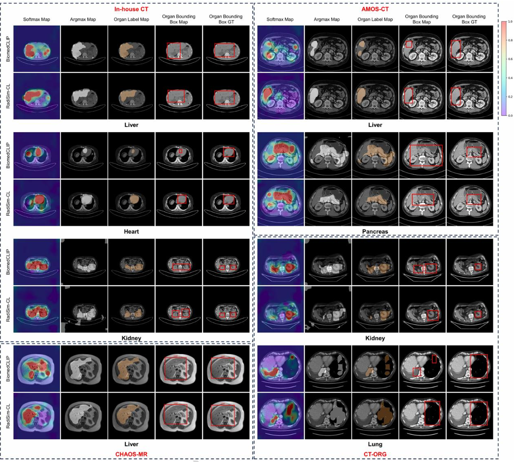
- Anatomical Localization: The model doesn't just "guess" a label; its attention maps align with expert-annotated boundaries of organs like the liver and kidneys.
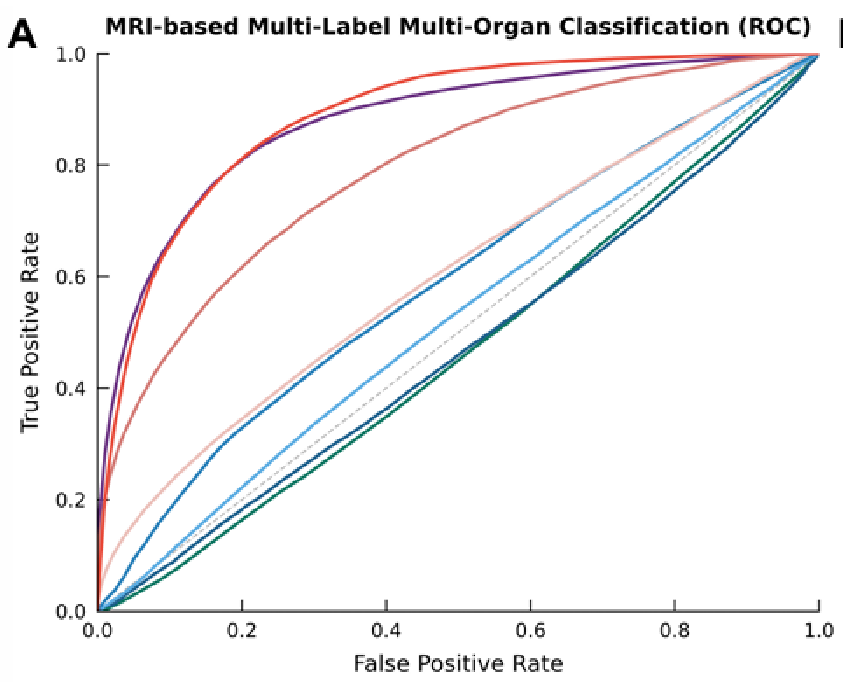
- Zero-Shot Diagnostic Superiority: In the challenging task of diagnosing brain tumors (Br35H dataset), RadiSim-CL achieved an AUC of 0.953.
- The Ultimate Test (Grading): Most models fail at intra-class variation. RadiSim-CL achieved 0.764 accuracy in Meningioma grading, nearly double the performance of specialized medical baselines like BiomedCLIP (0.349).
Deep Insight: Why Curriculum Learning?
An ablation study revealed a startling fact: a model trained on the same data but without the sequential curriculum (Uniform training) performed significantly worse on complex tasks. For example, in knee ACL tear detection, the uniform model was barely better than a coin flip, while RadiSim-CL excelled. This suggests that foundational knowledge acts as a scaffold—without a solid understanding of anatomy, the model cannot learn the "high-level semantics" required for diagnosis.
Critical Analysis & Future Outlook
While RadiSim-CL is a major leap toward "Radiologist-level" AI, the authors remain humble. The model still struggles with:
- Small organ confusion: Morphologically similar structures (like small vessels vs. nodules) can still cause errors.
- Temporal tracking: Real-world radiology often requires comparing current scans with historical ones to track disease progression—a "longitudinal" capability current VLMs still lack.
The Takeaway: RadiSim-CL proves that for AI to succeed in high-stakes fields like medicine, we must move beyond "big data" and toward "structured wisdom." By teaching AI the way we teach humans, we can build models that are not just accurate, but clinically logical.