
Bridging the Gap in Clinical Imaging: Machine Learning for Personalized Breast Cancer and Diabetes Care
Machine Learning in Healthcare: Breast Cancer and Diabetes Cases
This paper presents a comprehensive workflow of machine learning and image analysis applications in healthcare, specifically focused on personalized breast cancer screening and diabetes monitoring. The author introduces CASAM-Vol for volumetric breast density estimation and specialized algorithms like COM-AR for enhancing Optical Projection Tomography (OPT) in diabetes research.
TL;DR
In the evolving landscape of medical AI, the transition from research to clinical reality often hits a bottleneck: data compatibility and imaging artifacts. This paper presents a multidisciplinary framework that solves two major hurdles: estimating breast cancer risk from processed mammograms where standard tools fail, and automating the precision imaging of pancreatic cells in diabetes research. By leveraging "shallow" ML for calibration and Deep Learning for diagnostics, the study provides a blueprint for evidence-based personalized medicine.
Problem & Motivation: The Reality of "Lost" Data
The clinical environment is often a graveyard for high-fidelity raw data. Hospitals typically store "For Presentation" mammograms (processed for human viewing) and discard the "Raw" data required by gold-standard volumetric tools like Volpara. This creates a gap where advanced risk assessment is impossible for historical or standard processed archives.
In the realm of diabetes, Optical Projection Tomography (OPT) allows for 3D visualization of the pancreas, but it is hypersensitive to human error. If a specimen isn't perfectly centered on the Optical Axis of Rotation (OAR), the resulting 3D reconstruction is blurred, rendering Beta-cell quantification—the key to understanding diabetes progression—unreliable.
Methodology: Mimicking Experts and Correcting Physics
The author addresses these challenges through three distinct technological nodes:
1. CASAM-Vol: Mimicking Proprietary Algorithms
To bypass the need for raw FFDM data, the author developed CASAM-Vol. This tool uses a Random Forest regressor trained on a combination of statistical/morphological features and DICOM header tags (like KVP and Exposure) to predict what the Volpara software would have calculated from the raw data.
2. COM-AR: Precision Bioimaging
For OPT, the author introduced Center of Mass based Axis Rotation (COM-AR). Utilizing the Expectation-Maximization (EM) algorithm, this tool calculates the required displacement to align a specimen to the OAR post-scan, effectively "fixing" manual mounting errors in software.
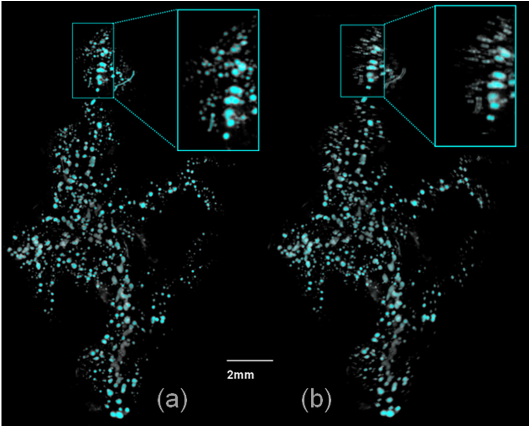 Fig 1: Volume rendering of a specimen using the proposed approach (a) vs the variance-based commercial package (b).
3. Deep Learning for Retinopathy
The approach to Diabetic Retinopathy moves beyond simple classification. By segmenting the fundus image into the optic disc, blood vessels, and "other" regions, the research found that the "other" regions (often containing hard exudates) actually hold more predictive power than the full image when fed into architectures like AlexNet and ResNet50.
Experiments & Results: Quantitative Validation
The effectiveness of these methods was validated across substantial datasets:
- Breast Cancer Risk: CASAM-Vol was tested on 47 cases and 1011 controls. All measures, including the "mimicked" volumetric density, showed a statistically significant association with breast cancer risk and specific genetic variants (rs10995190).
- Pectoral Muscle Insight: A surprising finding was that the pectoral muscle—usually discarded in mammogram analysis—showed a significant association with cancer risk even after adjusting for age and BMI.
- OPT Enhancement: The tool effectively removed y-axis shifts using Fourier transforms and linear regression, providing much clearer visualization of insulin-producing islets.
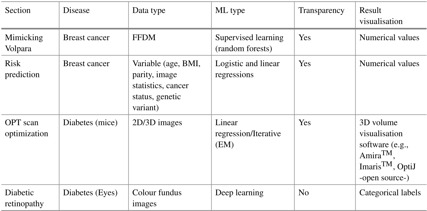 Table 1: Summary of ML types and data applications used in the study.
Critical Analysis & Conclusion
Takeaway
The paper proves that ML doesn't always need to be a "black box." In the cases of breast density and OPT centering, ML acts as a bridge, translating processed clinical data back into a format that provides quantitative biological insights.
Limitations
While the breast cancer models are robust, the CASAM-Vol tool is currently limited to specific manufacturers (GE, Sectra, Philips) and requires extensive DICOM acquisition parameters to be present in the metadata. If these tags are missing, the volumetric estimation cannot proceed.
Future Outlook
The shift towards personalized medicine depends on our ability to extract "hidden" biomarkers. Whether it is tissue stiffness or pectoral muscle attenuation, the future of healthcare AI lies in identifying features that the human eye ignores but the algorithm can quantify.