
M.tb IDH-PPARγ Interaction: A Metabolic Hijacking that Drives Macrophage Ferroptosis
Mycobacterium tuberculosis IDH-PPARγ interaction suppresses GPX4 to drive macrophage ferroptosis and sustain persistent infection
This study identifies a novel mechanism where Mycobacterium tuberculosis (M.tb) isocitrate dehydrogenase (IDH) interacts with host PPARγ to drive macrophage ferroptosis. By stabilizing PPARγ and recruitment of the NCOR/SMRT corepressor complex, M.tb suppresses the antioxidant enzyme GPX4, facilitating bacterial persistence and lung inflammation.
TL;DR
Mycobacterium tuberculosis (M.tb) doesn't just hide in macrophages; it actively rewires their metabolic circuitry to ensure its own survival. This study reveals that M.tb releases isocitrate dehydrogenase (IDH), which "hijacks" the host protein PPARγ. This interaction prevents the degradation of PPARγ, causing it to accumulate and repress the vital antioxidant enzyme GPX4. The result? A surge in lipid peroxidation and ferroptosis, which destroys the macrophage and allows the bacteria to spread.
Problem & Motivation: The Mystery of Persistent Infection
M.tb is a master of persistence. It is well-known that the pathogen reprograms host lipid metabolism, turning macrophages into "foamy cells" filled with lipid droplets. However, the exact molecular link between this lipid accumulation and the eventual death of the host cell via ferroptosis remained a "black box." Existing research pointed toward GPX4 depletion, but the why and how of this depletion—specifically which bacterial proteins were pulling the strings—remained unknown.
Methodology - The Core: Identifying the IDH-PPARγ Axis
The researchers utilized a multi-omics approach—combining single-cell RNA sequencing (scRNA-seq) of TB patient samples with proteomics—to pinpoint PPARγ as a key player that is upregulated during infection.
1. The Hijacking Mechanism
Through Co-Immunoprecipitation (Co-IP) and structural modeling, the authors identified that M.tb IDH (Rv3339c) interacts directly with PPARγ. This interaction is sophisticated: it physically blocks the S112 phosphorylation site on PPARγ, a modification that normally signals the protein for degradation via the Ubiquitin-Proteasome System (UPS).
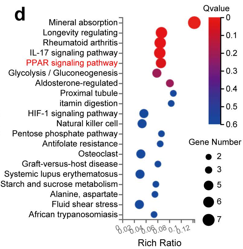 Figure: Predicted binding model of mouse PPARγ (orange) and M.tb IDH (marine), showing the stabilization of the host protein.
2. Transcriptional Repression of GPX4
Once stabilized, the "excess" PPARγ translocates to the nucleus. Instead of acting as a typical activator, it recruits the NCOR/SMRT corepressor complex to the promoter region of the Gpx4 gene. This recruitment leads to decreased chromatin accessibility and the silencing of Gpx4 expression, stripping the cell of its primary defense against lipid peroxidation.
Experiments & Results: Validating the Axis
The study utilized both in vitro macrophage models and in vivo mouse models (including tamoxifen-inducible PPARγ knockout mice) to prove the functional impact of this axis.
Key Findings:
- Bacterial Load: Inhibiting PPARγ with the antagonist GW9662 or through genetic knockout significantly reduced the bacterial burden in the lungs.
- Ferroptosis Markers: M.tb infection led to a massive increase in FerroOrange (iron) and C11 BODIPY (lipid peroxidation) signals, both of which were reversed when the PPARγ-GPX4 axis was restored.
- Mitochondrial Health: TEM imaging showed that PPARγ inhibition partially restored mitochondrial structure, which is typically shrunken and damaged during ferroptosis.
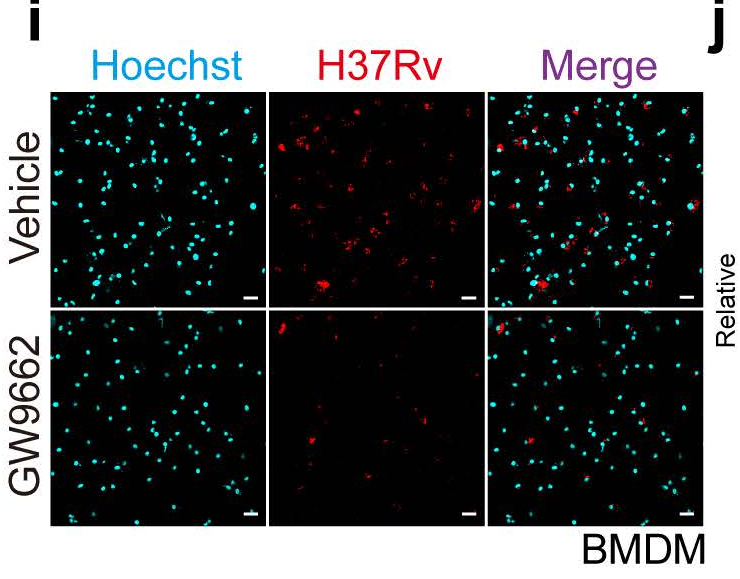 Figure: Experimental evidence showing that hindering the PPARγ axis reduces cell death and lipid peroxidation.
Critical Analysis & Conclusion
Takeaway
The discovery of the IDH-PPARγ-GPX4 axis provides a clear roadmap for Host-Directed Therapy (HDT) in TB. By targeting a host pathway rather than the bacteria directly, researchers can bypass the growing problem of multi-drug resistant (MDR) TB.
Limitations & Future Outlook
While the study provides robust evidence for IDH's role, the authors note it remains unclear exactly how a non-classically secreted protein like IDH escapes the bacterial cell to interact with the host cytosol—hypothesizing bacterial lysis or ESX-1-driven membrane disruption. Furthermore, the findings raise a significant red flag for diabetic patients: the common use of PPARγ-agonist drugs (like TZDs) for blood sugar control might inadvertently exacerbate TB infections by facilitating this ferroptotic pathway.
In conclusion, M.tb's ability to turn a host's metabolic regulator (PPARγ) into a weapon against itself (GPX4 repression) is a testament to the evolutionary complexity of this ancient pathogen. Restoring this balance may be the key to finally eradicating persistent TB.