
Empowering Liver Surgery: A Feature-Driven 3D Ultrasound Visualization Breakthrough
Development of novel algorithm to visualize blood vessels on 3D ultrasound images during liver surgery
The paper introduces a novel volume visualization algorithm specifically designed for 3D B-mode ultrasound images in liver surgery. By replacing traditional transfer functions (TF) with a feature-based importance mapping, the method achieves high-resolution visualization of hepatic blood vessels and tumors, aiding surgeons in avoiding vascular injury during operations.
TL;DR
Navigating the complex vascular network of the liver during surgery is a high-stakes task where a single misstep can lead to catastrophic hemorrhaging. This paper presents a novel algorithm that transforms noisy 2D B-mode ultrasound slices into high-fidelity 3D volume renderings. By moving away from cumbersome "Transfer Functions" and utilizing a specific blend of vessel-enhancing features (GVF and Frangi filters), the authors provide surgeons with a clearer, interactive map of the patient's internal anatomy in real-time.
The "Shadow" Problem in Clinical Ultrasound
While CT and MRI provide crisp 3D views, they aren't practical for real-time adjustments in the operating room. Ultrasound is portable and non-invasive, but its 3D visualization has long been plagued by Speckle Noise and Shadow Artifacts.
Previous attempts to solve this used 1D Transfer Functions (mapping intensity to color), which often confuse noise with tissue, or Multi-Dimensional Transfer Functions (MDTF), which are so mathematically complex that clinicians find them too difficult to "tune" during a surgery. The result? Most surgeons revert to 2D images, losing the vital spatial context of how vessels curve through the liver.
Methodology: Beyond the Transfer Function
The core innovation lies in the Classification Step. Instead of a global map, the algorithm treats every voxel as a carrier of specific biological features.
1. Feature Extraction Pipeline
The system extracts three primary characteristics to identify "Areas of Interest":
- Gradient Vector Flow (GVF): Used to capture object boundaries and tubular structures (vessels) while ignoring noise.
- Frangi Filter (Vesselness): Specifically amplifies the look of blood vessels by analyzing the Hessian matrix eigenvalues.
- Sobel Gradient: Provides the fundamental structural edges of the liver and potential tumors.
2. Importance Mapping and HSL Coloring
The algorithm introduces an "Importance Degree" (). A surgeon can adjust the importance of "Vessels" vs. "Tissue" through a simple slider. These inputs are processed through an adaptive opacity formula:
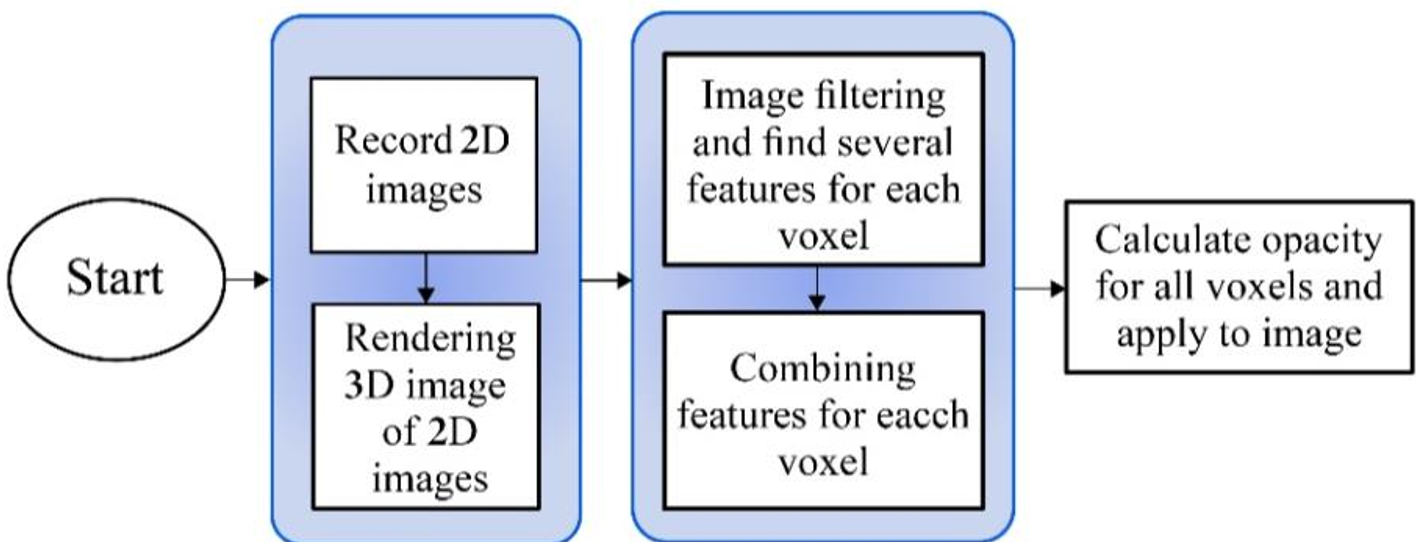
Figure 1: The schematic flow—from 2D acquisition to voxel-wise feature calculation and final 3D rendering.
Experimental Results: Seeing Through the Noise
The team tested the algorithm on real-world datasets from patients at Imam Khomeini Hospital. The results were quantified using Peak Signal-to-Noise Ratio (PSNR) and Mean Square Error (MSE), but the true value was in the visual clarity.
- Noise Suppression: Fast bilateral filtering removed speckle noise (PSNR ~28dB) without blurring the critical vessel edges.
- Vessel Delineation: Compared to the standard Maximum Intensity Projection (MIP), the new method allowed the Frangi and GVF signatures to "pop," showing small bifurcations that were previously invisible.
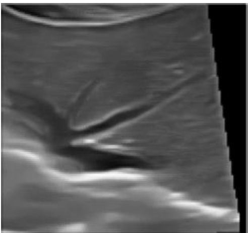
Figure 2: The bottom image shows the enhanced clarity achieved by combining all three algorithms, compared to single-feature importance.
Critical Insight: Why This Matters
The shift from Intensity-based rendering to Feature-based rendering is a paradigm shift. In B-mode ultrasound, a pixel's "brightness" (intensity) is a poor indicator of what it actually is (it could be a vessel wall, a tumor, or just a noise artifact). By using geometric features like "vesselness," the algorithm provides a semantic layer to the visualization.
Limitations & Future Outlook
While the method is a significant step forward, some hurdles remain:
- Rib Shadows: Acoustic shadows from bones can still create "dead zones" in the 3D volume.
- Human-in-the-loop: The system still requires a clinician to set initial importance weights, though this is much simpler than tuning an MDTF.
The authors suggest that the next frontier is Deep Learning, where neural networks could automatically identify these features, further reducing the cognitive load on the surgeon.
Conclusion
This novel algorithm successfully bridges the gap between complex computer graphics andpractical clinical utility. By providing high-quality, 3D spatial awareness of the liver's vascular "tree," it offers a direct path to safer, more precise surgeries.