
EARNN: Interpretable Brain Disorder Prediction via Regionalized Deep Learning
A novel ensemble approach on regionalized neural networks for brain disorder prediction
EARNN (Ensemble Approach on Regionalized Neural Networks) is a novel two-stage deep learning framework for brain disorder prediction using fMRI data. It leverages 3D Convolutional Neural Networks (CNNs) and a unique stochastic pooling mechanism to extract regional biomarkers, achieving state-of-the-art performance on HIV and ADHD datasets.
TL;DR
Predicting brain disorders like ADHD and HIV from fMRI data is notoriously difficult due to the high dimensionality and non-linear nature of neural connectivity. EARNN (Ensemble Approach on Regionalized Neural Networks) tackles this by decomposing the brain into functional regions, training specialized 3D CNNs for each, and ensembling them via an EM algorithm. This method doesn't just improve accuracy—it tells doctors which parts of the brain are likely damaged.
Problem & Motivation: The "Black Box" of Whole-Brain Analysis
Most current neuroimaging analysis suffers from two extremes:
- Hand-crafted features: Methods like SVM or Logistic Regression treat voxels as independent variables, completely ignoring the spatial "structural information" of the brain.
- Global Deep Learning: Standard CNNs process the whole brain image uniformly. While they capture non-linear patterns, they ignore the biological reality that different diseases (e.g., Alzheimer's vs. ADHD) target very specific Regions of Interest (ROIs).
The authors' insight was simple yet profound: If diseases are regional, our architecture should be regional.
Methodology: The Architecture of EARNN
The EARNN framework operates in two distinct stages.
Stage 1: The Regionalized CNN (CNNr)
Instead of one giant model, the authors create individual networks, where each network is specialized for a specific brain region .
The secret sauce is the Regionalized Stochastic Pooling. Traditional pooling (Max or Average) just looks for the strongest signal. EARNN’s pooling uses a multinomial distribution where the probability of selecting an activation is weighted by whether that voxel actually belongs to the target region according to a brain atlas.
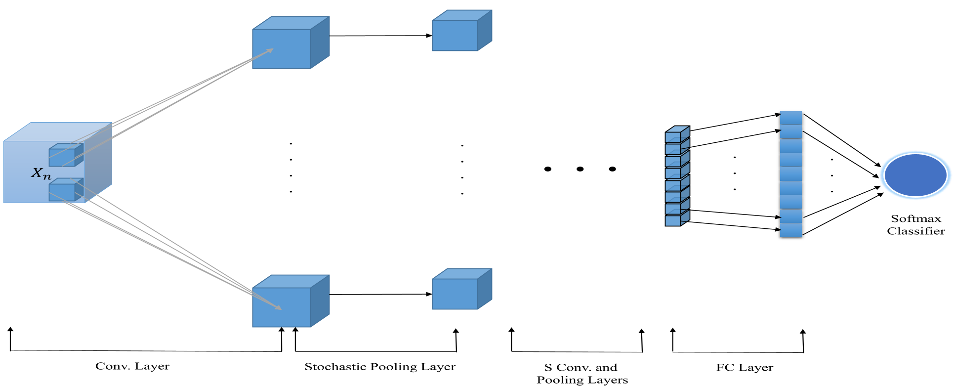 Figure 1: The architecture of a single regionalized CNN model showing the 3D convolution and the custom pooling layer.
Stage 2: EM-based Ensemble
Not all regions are equally important for every disease. EARNN uses an Expectation-Maximization (EM) algorithm to learn a set of mixing coefficients .
- E-Step: Estimate the "responsibility" of each regional network for the correct classification of a sample.
- M-Step: Update the weights to favor the regions that provide the most reliable diagnostic signals.
Experiments & Results: Accuracy Meets Interpretability
The model was tested on two challenging datasets: HIV (Northwestern University) and ADHD-200.
Quantitative SOTA Performance
EARNN-2 (a 2-layer variant) consistently outperformed all baselines. In the ADHD dataset, traditional methods like SVM hovered around 45-54% accuracy (barely better than a coin flip), while EARNN achieved 76.5%.
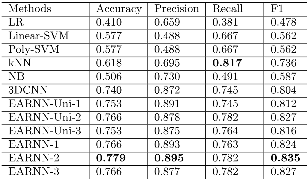 Table 1: Performance comparison on the HIV dataset. EARNN shows a clear lead in F1 score and Precision.
Identifying the "Target" of Disease
Perhaps the most significant result is shown in the mixing coefficients. By ranking the highest values, EARNN identified regions like the Temporal Superior Gyrus and Precuneus as high-impact areas for HIV and ADHD respectively—findings that align perfectly with established clinical domain knowledge.
 Table 2: Top brain regions identified by EARNN as relevant to HIV and ADHD.
Critical Analysis & Conclusion
Takeaway
EARNN proves that incorporating anatomical priors (via atlases and regional pooling) is superior to "blind" end-to-end learning for medical imaging. It effectively handles the structural complexity of fMRI while remaining interpretable.
Limitations
While powerful, the model relies on a pre-defined brain atlas. If the atlas is inaccurate or if the disease causes significant structural shifts that mismatch the atlas, the regional pooling might focus on the wrong voxels. Furthermore, the training of separate CNNs can be computationally expensive compared to a single sparse model.
Future Work
The next logical step for this research is moving from static fMRI to Dynamic Functional Connectivity, where the temporal changes in regional influence are modeled over the course of a scan.