
Seizure Detection: Boosting Epilepsy Diagnostics via XGBoost and PCA
Predictive analytics in healthcare epileptic seizure recognition
2018-10-29
Summary
Problem
Method
Results
Takeaways
Abstract
This paper presents a machine learning framework for the automated recognition of epileptic seizures using EEG data. By leveraging a multi-class dataset from the UCI repository, the authors evaluate several supervised learning algorithms, achieving high-performance SOTA for seizure detection using XGBoost for binary classification and Random Forest/MLP for multinomial classification.
## Executive Summary
**TL;DR**: This research tackles the unpredictability of epileptic seizures by converting raw EEG voltage fluctuations into a robust predictive model. By employing **XGBoost** and **Multi-Layer Perceptrons (MLP)**, the authors achieved an impressive **98.1% accuracy** in distinguishing seizure activity from healthy brain states, proving that machine learning can effectively substantiate clinical diagnoses with minimal human intervention.
**Academic Positioning**: This work acts as a bridge between traditional signal processing and modern ensemble learning. It moves beyond simple binary "Seizure vs. No Seizure" models to explore a 5-class spectrum (including tumorous zones and different wakeful states), identifying which clinical parameters are truly essential for diagnostic accuracy.
## The Core Challenge: Finding the Signal in the Noise
Interpreting EEG data is notoriously complex. Brain waves (Delta, Theta, Alpha, Beta) are unique to individuals and vary wildly based on activity—simply closing one's eyes can drastically shift the signal. Traditionally, diagnosing epilepsy required the "Ictal" state (the seizure itself) to be recorded, which is time-consuming and labor-intensive.
The authors asked: *Can we predict the probability of a seizure using parameters from non-seizure periods?* This would allow for smarter, faster, and cheaper diagnostic tools.
## Methodology: From Raw Voltage to Predictive Power
The study utilized a dataset of 11,500 instances, each containing 178 features of digitized voltage signals.
### 1. Data Structuring and Class Discovery
The researchers didn't just accept the labels; they used **Hierarchical Clustering (WARD method)** to verify that the data naturally organized into five distinct clinical conditions, ranging from "Eyes Open/Healthy" to "Seizure Activity."
### 2. Dimensionality Reduction (PCA)
To handle the high feature count (178 attributes), the team employed **Principal Component Analysis (PCA)**. They found that the first 50 components captured the vast majority of variance, allowing classifiers to run more efficiently without losing diagnostic integrity.
### 3. Machine Learning Architecture
- **Binary Classification**: Seizure (Class 1) vs. All others.
- **Multinomial Classification**: Distinguishing between all five categories simultaneously.
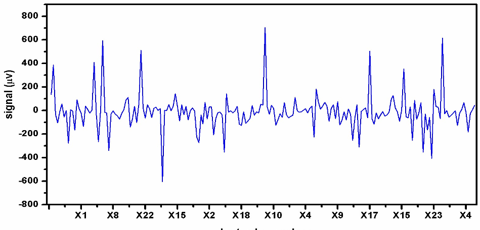
*Figure 1: Digital regeneration of analog EEG signals from 23 electrodes.*
## Results: The Power of Boosting
The experimental results highlighted a clear winner in the binary task: **XGBoost**.
| Classifier | Accuracy | F-score |
| :--- | :--- | :--- |
| **XGBoost (Binary)** | **98.1%** | **98.5%** |
| **SVC (Binary)** | 97.17% | 96.7% |
| **MLP (Multinomial)** | 71.32% | 93.9% (Class 1) |
| **Random Forest** | 75.69% | - |
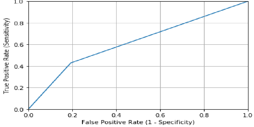
*Figure 2: ROC curves showing the high sensitivity and specificity of the model in distinguishing seizure states.*
### Key Insight: The Redundancy of Tumorous Zone Probing
One of the most profound findings was that probing the specific **tumorous zone (Class 2)** was actually a *weaker* predictor of seizures than probing the healthy part of the brain on the opposite hemisphere **(Class 3)**. This suggests that the "background" activity of a brain predisposed to epilepsy is more informative than the localized signal of a tumor.
## Critical Analysis & Conclusion
**Takeaway**: The study proves that ensemble methods like XGBoost are superior for EEG analysis because they handle the non-Gaussian distribution of voltage signals better than traditional logistic regression or k-NN models.
**Limitations**: While the results are high, the data is derived from a single (though reputable) source. Cross-dataset validation is necessary to ensure these features aren't overfitted to the specific recording equipment used in the UCI study.
**Future Outlook**: The next logical step is integrating these models into wearable EEG "patches" that can alert patients or doctors to seizure risks in real-time, effectively moving epilepsy management from reactive treatment to proactive prevention.