
Probabilistic MS Lesion Classification: Why Local Context and Regional Variance Matter
9991_Probabilistic Multiple Sclerosis Lesion Classification Based on Modeling Regional Intensity Variability and Local Neighborhood Information.
This paper presents a fully automatic probabilistic framework for classifying Multiple Sclerosis (MS) lesions (T1-hypointense and T2-hyperintense) and healthy brain tissues. The method integrates regional intensity variability through lobe-specific likelihood modeling and local spatial context using Markov Random Fields (MRFs), achieving SOTA performance on multisite clinical datasets.
TL;DR
Researchers have developed a fully automated probabilistic framework that tackles the "intensity inconsistency" problem in Multiple Sclerosis (MS) MRI analysis. By dividing the brain into anatomical regions and applying Markov Random Fields (MRFs), the system significantly outperforms global intensity classifiers, particularly in difficult areas like the posterior fossa and for capturing small, low-contrast lesions.
The Problem: The "Moving Target" of MRI Intensities
Multiple Sclerosis diagnosis depends heavily on identifying lesions in White Matter. However, automated systems face three major hurdles:
- Regional Variability: A lesion in the frontal lobe looks different from a lesion in the posterior fossa.
- Multisite Noise: Different MRI scanners produce different intensity scales.
- Boundary Ambiguity: "Dirty white matter" around lesions creates fuzzy edges that lead to high false-positive rates.
Most prior work treated the entire brain as a single intensity distribution or relied on voxel-specific templates that break down if registration is slightly off.
Methodology: Region-Specific Likelihoods & MRF Smoothing
The core innovation lies in the Hierarchical Probabilistic Model. Instead of one global model, the authors segment the brain into six regions (Frontal, Parietal, Temporal, Occipital lobes, Central region, and Posterior Fossa).
1. Regional Likelihoods
Within each region , the system models each tissue class (WM, GM, CSF, T1-lesion, T2-lesion) as a multivariate Gaussian. This accounts for the physical fact that tissue properties are not uniform across the organ.
2. The Power of MRFs
To prevent "salt and pepper" noise (isolated misclassified pixels), the authors use Markov Random Fields. They don't just look at single-voxel cliques but use 2, 5, and 7-voxel cliques to capture the "blob-like" nature of lesions.
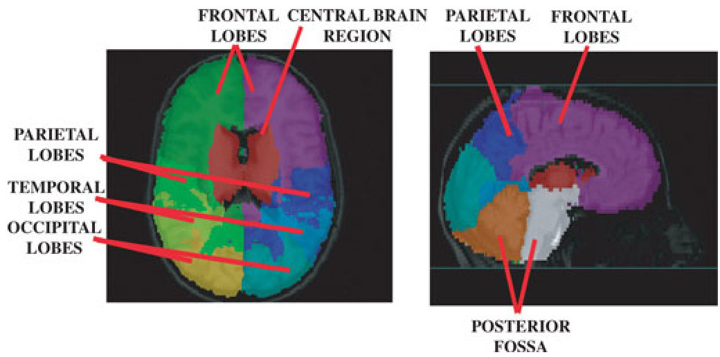 Figure 1: The two-stage workflow showing Training (Atlas and Likelihood building) and Classification (MAP initialization followed by ICM refinement).
Experiments & SOTA Results
The model was tested on a robust dataset of 90 multisite patients.
- The Posterior Fossa Win: This region is notoriously difficult. The proposed method achieved a Dice Kappa of 0.37, a huge leap over the 0.22 achieved by standard outlier-detection methods.
- Sensitivity: Voxel-based sensitivity reached 0.784, outperforming the widely used Van Leemput method.
- Uncertainty Quantification: By calculating Shannon's Entropy on the posterior distribution, the system provides a "confidence map." This allows clinicians to focus only on voxels where the algorithm is "unsure."
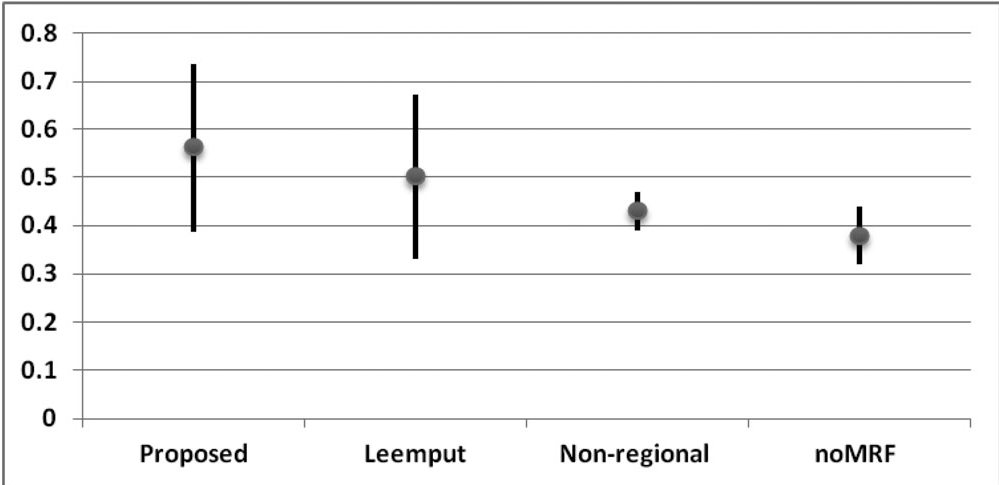 Figure 2: Qualitative comparison. Our method (a) shows significantly fewer false negatives (green) and false positives (blue) compared to non-regional methods (c) or those without MRF (d).
Critical Insight: The Value of "Structured" Probability
While modern Deep Learning (CNNs/Transformers) has largely taken over segmentation, this paper provides a timeless lesson: Domain-specific structural priors (like anatomical regions) are powerful regularizers.
The transition from global statistics to region-based statistics mirrors the "Attention" mechanisms in modern AI—focusing the model's "expectations" based on the spatial context. For practitioners today, the use of Entropy as a filter for post-processing remains a highly effective way to reduce the workload of expert raters in clinical trials.
Conclusion
This work demonstrates that by acknowledging the brain's inherent anatomical variability and using MRFs to enforce local consistency, we can create automated tools that rival human experts. The success in multisite trials proves that "regional intelligence" is the key to handling real-world clinical noise.