
Dynamic Scan Parameter Optimization for CT-Guided Interventions: Beyond the 80kV Myth
Towards Prospective Identification of Optimal Scan Parameters for CT-guided Interventions -A Phantom Study
This study evaluates how CT tube voltage and tube load affect the Contrast-to-Noise Ratio (CNR) in CT-guided interventions across varying patient sizes and target tissues. Using a modular multi-energy phantom (20 cm and 35 cm), the authors established tailored scan protocols that optimize image quality relative to radiation dose.
TL;DR
This phantom study debunked the universal recommendation of low tube voltage (kV) for CT-guided interventions. While 80 kV excels for iodine targets in pediatric-sized phantoms, it drastically fails in adult-sized patients (35 cm diameter) where 140 kV becomes the superior choice for maximizing the Contrast-to-Noise Ratio (CNR). The research provides a definitive roadmap for adaptive protocol selection based on target tissue and patient body diameter.
Problem & Motivation: The Failure of "One-Size-Fits-All"
In interventional radiology, the primary goal is the clear identification of a Tissue of Interest (TOI). Traditionally, clinicians have favored low-kV (typically 80 or 100 kV) protocols, operating under the assumption that lower energy increases tissue attenuation and thus image contrast.
However, the authors point out a critical flaw: image quality is a function of both contrast and noise. Lowering the tube voltage exponentially increases image noise and reduces beam penetration, especially in larger patients. This trade-off often leads to "grainy" images where the target is lost in the noise, necessitating repeat scans and increasing the total radiation dose. The core motivation of this study was to identify exactly when the benefits of low kV are negated by the penalty of increased noise.
Methodology: A Modular Phantom Approach
The researchers employed a modular multi-energy phantom capable of simulating two patient profiles:
- Small (20 cm): Representing a pediatric or slim adult patient.
- Large (35 cm): Representing a standard adult patient.
They tested three distinct target rods: Iodine-blood mixture (high contrast), Pure Calcium (high contrast), and Pure Blood (low contrast/soft tissue). By systematically varying tube voltages from 80 kV to 140 kV and adjusting mAs to maintain equivalent Size-Specific Dose Estimates (SSDE), they isolated the effect of photon energy on the resulting CNR.
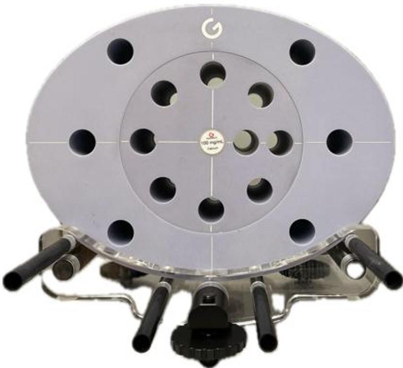 Fig 1. The multi-energy phantom setup used to simulate tissue types and patient diameters.
Results: The kV/Diameter Paradox
The study revealed a fascinating reversal of trends based on phantom size:
1. Small Phantom (20 cm)
- High Contrast (Iodine): 80 kV remains the "gold standard," providing the highest CNR due to the photoelectric effect's dominance near the iodine K-edge.
- Low Contrast (Soft Tissue): Higher voltages (140 kV) outperformed lower settings. The reduction in noise at 140 kV outweighed the marginal contrast loss in soft tissues.
2. Large Phantom (35 cm)
- Universal Failure of 80 kV: Regardless of the target tissue, 80 kV produced the lowest CNR. The limited beam penetration in the thicker phantom rendered low-energy photons ineffective.
- The High-Energy Advantage: For iodine targets at low doses (≤ 3 mGy), 140 kV was actually superior. As the dose increased, 120 kV became the optimal "sweet spot."
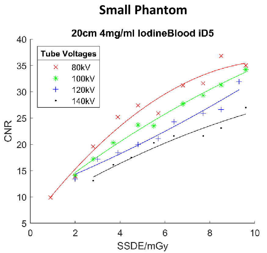 Fig 2. CNR curves showing the performance of different kV settings across dose levels. Note how 140 kV (blue line) dominates in larger diameters.
The Role of Iterative Reconstruction (IR)
While IR (iDose5) increased the overall CNR by approximately 34% through sophisticated denoising, it did not alter the fundamental relationship between tube voltage and tissue contrast. IR acts as an "amplifier" for image quality but cannot compensate for fundamentally poor parameter selection.
Critical Analysis & Conclusion
The study's most significant contribution is the clinical decision matrix derived from the data. It shifts the paradigm from "dose reduction at all costs" to "CNR optimization through adaptive physics."
| Tissue of Interest | Small Diameter (~20cm) | Large Diameter (~35cm) |
|---|---|---|
| Soft Tissue | 140 kV | 140 kV |
| Calcium | 100 kV | 140 kV |
| Iodine-Blood | 80 kV | 140 kV (low dose) / 120 kV (high dose) |
Limitations: The study utilized water-equivalent phantoms, which may not perfectly replicate human beam-hardening effects from bone or specialized tissue types. Furthermore, the absence of metallic interventional tools (needles) means the impact of spectral metal artifacts at different kV levels remains a topic for future research.
Takeaway: For modern CT-guided interventions, radiologists should move away from standard 80-100 kV protocols for adult patients, as 140 kV typically provides the superior balance of penetration and noise reduction needed for accurate target identification.