
Digitizing the Slit-Lamp: Empowering Rural Health Workers in Cataract Diagnosis
An interface to aid rural health workers in the preliminary diagnosis of cataract at the slit lamp using LOCS III
This paper presents a digital interactive interface designed to assist rural health workers in India with the preliminary diagnosis and grading of cataracts. By utilizing the Lens Opacities Classification System III (LOCS III) through a computer-based visual comparison tool, the system enables non-specialists to perform screenings that typically require an ophthalmologist.
TL;DR
With a massive shortage of eye specialists in rural India, this paper proposes a digital interface that allows trained health workers to diagnose cataracts using the LOCS III standard. By transforming a manual, expert-driven comparison process into an intuitive, computer-aided visual task, the system aims to make eye care accessible and reduce unnecessary travel for the rural poor.
Background: The Rural Healthcare Gap
In India, nearly 75% of the population resides in rural villages, yet 75% of doctors are concentrated in urban centers. Cataracts account for roughly 77% of blindness in the country. The current bottleneck isn't just the surgery itself, but the screening process. Most patients must travel hours to a city only to find their cataract isn't "mature" enough for surgery, wasting precious time and money.
The Problem: The Complexity of LOCS III
The Lens Opacities Classification System III (LOCS III) is the gold standard for grading cataracts. It involves comparing a patient's eye under a slit lamp to a set of standardized photographs.
- Subjectivity: Specialists must interpolate between photos (e.g., deciding if a cataract is a 2.4 or a 2.5).
- Physical Constraints: Dealing with physical transparency sheets is cumbersome in field conditions.
- Expertise Bottleneck: Only highly trained ophthalmologists are typically trusted with this grading.
Methodology: Visual Comparison & Interaction Design
The author developed an interactive interface that bridges the gap between a novice worker and an expert diagnostic.
1. The Dynamic Comparison Model
Instead of static photos, the interface provides a range of gradually varying alternatives. When a health worker identifies two reference images that "bracket" the patient's condition, the system generates animated sub-images between those points.
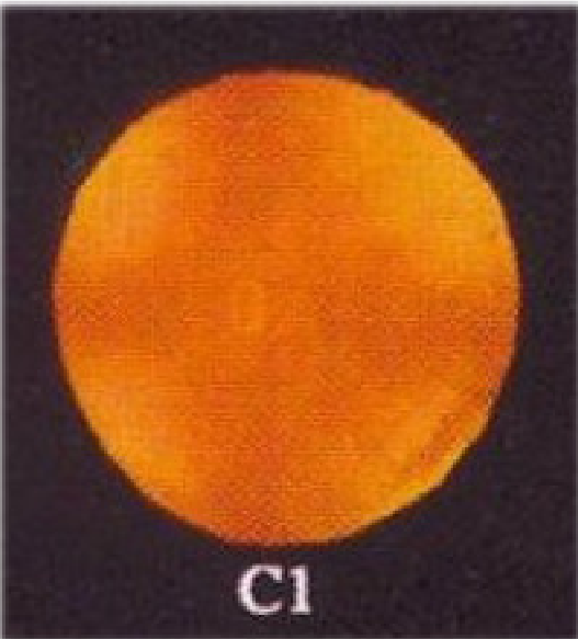 Figure 1: The UI allows the patient's image (left) to be compared against historical data and reference standards.
2. Progressive Animation
To prevent "user blindness" or confusion when images change, the system uses deliberate animations. When clicking a base image, sub-images "grow" out of it, providing a visual cue of the progression in severity.
 Figure 2: The zooming and selection process for fine-grained decimal grading.
Experimental Insights
During user testing at Shankar Netralaya Guwahati, several key insights emerged:
- Historical Comparison: Workers found it extremely valuable to see the image from the previous visit side-by-side with the current one to track the degradation rate.
- Animation Matters: Users preferred seeing how the image changed (progressive transitions) rather than sudden updates, which helped them understand the scale of severity.
Critical Analysis & Conclusion
This work represents an early and vital step in Telemedicine and ICT for Healthcare.
Takeaway: The success of the "ophthalmologist-led" model depends entirely on the reliability of the tools given to paramedics. By digitizing the LOCS III system, the author reduced the cognitive load of diagnosis, essentially moving the "intelligence" from the doctor's brain into the interface design.
Limitations: At the time of publication (2007), the system still required a manual slit-lamp setup and a computer. In today's context, this would likely be implemented as a mobile app utilizing AI/Computer Vision to automate the grading entirely. However, the human-centered design principles—specifically the focus on visual interpolation—remain highly relevant for medical UI today.
Future Outlook: Systems like this pave the way for massive, data-driven longitudinal studies on lens opacification, potentially revealing new insights into how cataracts progress in different demographic groups.