
Segmenting Lung Fields in Serial Radiographs: The Power of Patient-Specific Shape Statistics
Segmenting Lung Fields in Serial Chest Radiographs Using Both Population-Based and Patient-Specific Shape Statistics
This paper introduces a novel deformable model for lung field segmentation in serial chest radiographs by integrating population-based and patient-specific shape statistics. The method utilizes a modified SIFT descriptor for robust feature matching and an online learning framework to adapt the model to an individual's unique anatomy over time, achieving state-of-the-art accuracy in longitudinal medical imaging.
TL;DR
Lung field segmentation in chest X-rays is vital for calculating cardiac size and managing dialysis patients. However, standard "one-size-fits-all" models often fail to capture the subtle, unique anatomical changes in a single patient over time. This paper introduces a dynamically evolving deformable model that learns a patient's specific lung shape as new scans arrive, combined with SIFT descriptors for superior feature matching, pushing segmentation accuracy beyond 94%.
Problem & Motivation: Why General Models Fail
Traditional Active Shape Models (ASM) are like a "generic template." They work well for average cases but struggle with:
- Fuzzy Boundaries: The ribcage and diaphragm often have low contrast, leading "edge-seeking" algorithms astray.
- Visual Noise: Rib structures create repeated patterns that confuse simple gradient-based trackers.
- Anatomical Uniqueness: A population-based model contains the variance of everyone, which includes shapes impossible for a specific patient, while missing the specific nuances of that patient’s disease progression.
The authors' insight was simple but powerful: If we see the same patient every month, why not use their previous scans to build a personalized anatomical model?
Methodology: The Personalization Engine
The core innovation lies in the Energy Function (), which balances three forces:
- SIFT Feature Term: Unlike intensity gradients, SIFT (Scale-Invariant Feature Transform) provides a 128-dimensional "fingerprint" for each point on the lung boundary, making it much harder for the model to get "lost" in noise.
- Population Constraint (): Used initially to provide a "educated guess" based on a broad dataset.
- Patient-Specific Constraint (): An online, incrementally updated model.
The Online Learning Workflow
As the patient returns for scans (Time ), the system evolves:
- Phase 1 (): Rely on the Population model.
- Phase 2 (): Gradually "dial up" the patient-specific weight as more data is collected.
- Phase 3 (): Rely purely on the patient-specific model.
To handle the "small data" problem (modeling shape with only 5-10 images), the authors used Hierarchical PCA. Instead of modeling the whole lung at once, it breaks the contour into segments, capturing local variations more effectively than a global PCA could.
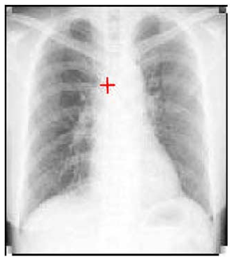 Figure 1: The incremental learning process for patient-specific segmentation.
Experiments & Results: Precision Through Personalization
The researchers tested their approach on the public JSRT database and a private serial radiograph dataset.
- Superior Logic: Using SIFT alone (ASM SIFT) improved the overlay percentage from 87% to 92% compared to standard intensity-based ASM.
- Personalization Leap: Adding patient-specific statistics pushed the accuracy to 94.9%.
- Stability: As shown in the comparisons below, the patient-specific methods (Method 1 & 2) provide significantly tighter and more consistent contours over months of scans.
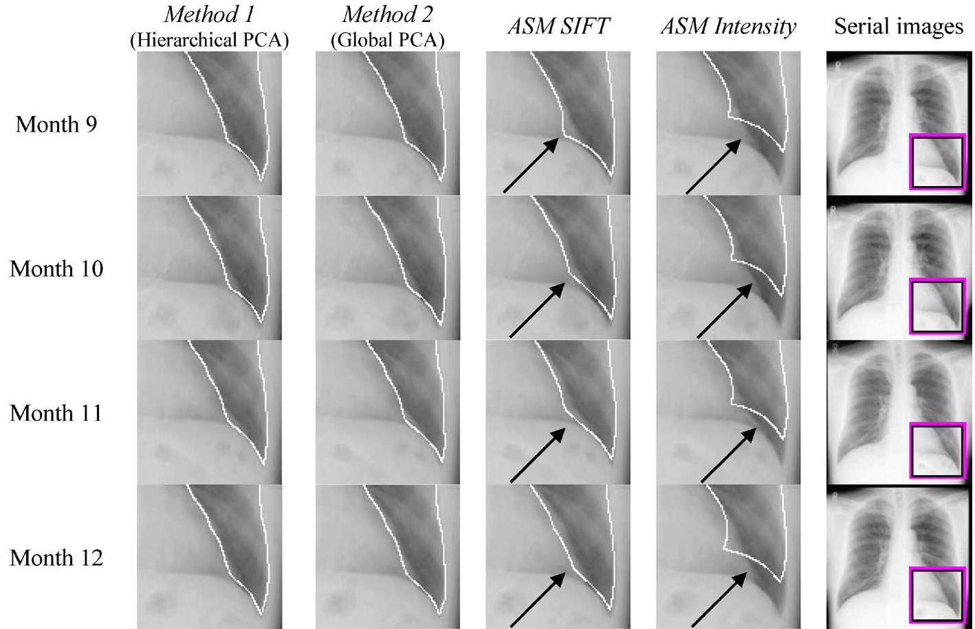 Figure 2: Comparison between standard ASM and the proposed personalized method. Black arrows point to errors in traditional methods corrected by the new approach.
The "Sweet Spot" of Learning
The study found that after 10 scans of a single patient, the model becomes highly specialized. At this point, the Hierarchical PCA and Global PCA converge in performance because the sample size finally suffices to capture the patient's individual anatomical manifold.
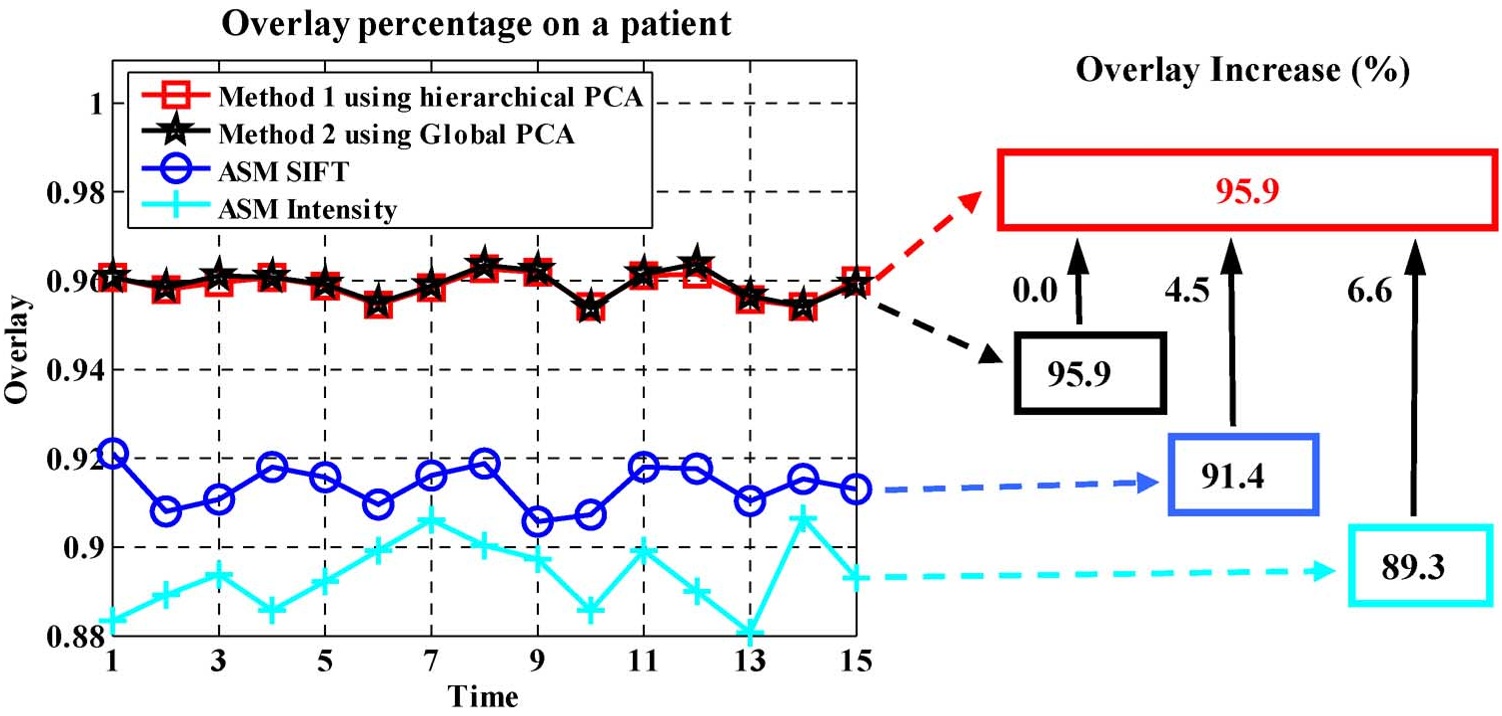 Figure 3: Accuracy improvement as more patient-specific data is acquired over time.
Critical Analysis & Conclusion
Why it Works
The success of this method stems from the Reduction of Search Space. By using a patient-specific model, the algorithm effectively says: "I know exactly what this patient's lung looks like; I just need to find its current pose." This is far more robust than asking: "Is this thing a lung?"
Limitations
- Computational Cost: SIFT descriptors are significantly slower to compute (roughly 2.5x slower than ASM Intensity).
- Initial Error Propagation: If the first few segmentations are wildly incorrect, the "patient-specific" model will learn those errors. User verification is currently required to ensure the system doesn't learn "bad habits."
Future Outlook
This paper serves as a precursor to modern Few-shot Learning and Personalized AI. In the future, we might see "Self-Correcting" models that use temporal consistency (the fact that lungs don't change shape drastically in 24 hours) to automatically discard poor segmentations without human intervention.