
Mapping the Malaria "Dark Proteome": A New Spatial Map of Plasmodium falciparum
The spatial proteome of the Plasmodium falciparum schizont illuminates the composition and evolutionary trajectories of its organelles
This study presents a high-resolution spatial proteome map of the Plasmodium falciparum schizont stage using hyperLOPIT. The researchers successfully resolved 24 distinct subcellular niches, classifying 1,646 proteins and providing the first comprehensive atlas of protein localization for the malaria parasite, including exported sites within the host cell.
TL;DR
Researchers have published a comprehensive spatial atlas of the Plasmodium falciparum schizont, the lethal stage of the malaria parasite. By employing hyperLOPIT spatial proteomics, they resolved 24 distinct cellular niches and localized over 1,600 proteins. This "GPS for proteins" reveals that the parasite's evolutionary energy is focused on the interface where it interacts with human red blood cells, providing a roadmap for future drug and vaccine development.
The Localization Crisis in Parasitology
Understanding Plasmodium falciparum is a game of biological hide-and-seek. Despite decades of research, a massive fraction of the parasite's proteome remains "uncharacterized"—we know the sequences, but we have no idea where they go or what they do.
This lack of context stems from two main issues:
- Divergence: Apicomplexans are so distantly related to yeast or humans that orthology (predicting function by similarity) often fails.
- Complex Architecture: The parasite resides inside a host erythrocyte, creating its own organelles and "exporting" proteins into the host cell. Mapping these shared and distinct compartments simultaneously is a technical nightmare.
Methodology: The hyperLOPIT Solution
The team used hyperplexed Localisation of Organelle Proteins by Isotopic Tagging (hyperLOPIT). Unlike traditional microscopy which looks at one protein at a time, hyperLOPIT uses density gradients and mass spectrometry to profile thousands of proteins simultaneously.
The core insight was the "Merozoite Refinement." Secreted proteins (like those in the rhoptries) often appear in two places: inside the organelle and at their final destination. By comparing schizonts (the parent stage) with purified merozoites (the daughter stage), the authors disentangled these signals, allowing for the high-resolution mapping of 24 compartments.
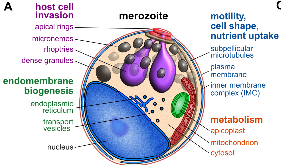 Figure 1: The hyperLOPIT workflow and the complex architecture of the infected erythrocyte.
Evolution and Adaptation: The "Subcellular Choreography"
By cross-referencing their spatial map with evolutionary data, the authors uncovered where the parasite is currently "innovating."
- Host-Interaction Sites: Compartments like the Maurer’s clefts and the Parasitophorous Vacuole Membrane (PVM) are hotspots for gene gain and positive selection.
- Metabolic Stability: In contrast, internal organelles like the mitochondria and apicoplast are under strict purifying selection, meaning their proteins rarely change because their functions are essential and fragile.
- Disorder as a Shield: Proteins exported to the host cell show high levels of intrinsic disorder, a potential strategy for immune evasion and flexible protein-protein interactions.
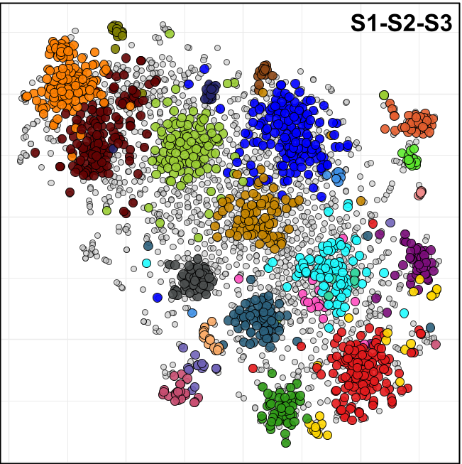 Figure 2: t-SNE projection showing the distinct clusters of proteins corresponding to 24 subcellular niches.
Experimental Validation
To ensure the machine learning wasn't hallucinating protein locations, the authors selected 15 uncharacterized proteins and used C-terminal HA-epitope tagging. In all cases where a signal was detectable, the microscopy (IFA) confirmed the hyperLOPIT prediction.
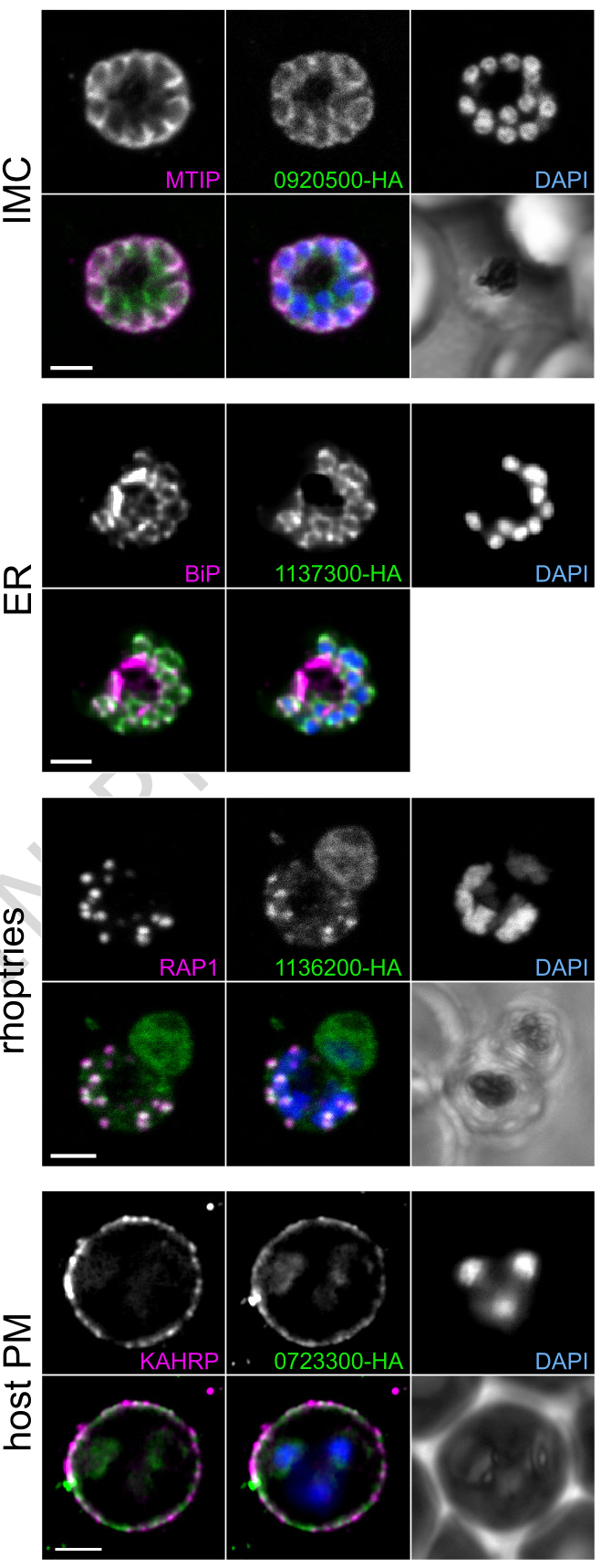 Figure 3: Immunofluorescence validation of uncharacterized proteins localized to specific organelles like the IMC and Rhoptries.
Critical Insight: The Apicoplast Paradox
The study highlights the Apicoplast—a remnant plant-like plastid—as a particularly interesting target. While it only produces one essential metabolite (isoprenoids) in the blood stage, nearly all its proteins are "essential" based on mutagenesis scores. This suggests the organelle's role is more integrated and structurally vital than previously thought, reinforcing its status as a primary drug target.
Conclusion & Future Outlook
This work transforms P. falciparum from a list of genes into a structured cellular machine. By providing the specific "address" for over 1,600 proteins, the researchers have created a resource that will allow others to prioritize targets for drug development.
Limitations: The study is a "steady-state" snapshot of the schizont stage. Many proteins are dynamic and may change locations during different phases of the lifecycle (e.g., ring or trophozoite stages). Future work will likely involve temporal spatial proteomics to capture the parasite in motion.
Takeaway: Innovation in malaria happens at the edges—the compartments that touch the host are the most diverse, the most disordered, and the fastest-evolving.