
Decoding Schizophrenia: High-Precision EEG Classification via Emotional Arousal
Specificity Analysis of Picture-Induced Emotional EEG for Discrimination Between Schizophrenic and Control Participants
This study presents a machine learning framework for discriminating between schizophrenia patients and healthy controls using picture-induced emotional EEG signals. By analyzing time-domain dynamics and hemispheric asymmetry, the authors achieved a peak F1-score of 0.994 using an SVM classifier with grid search optimization.
TL;DR
Researchers have developed a highly accurate (F1-score 0.994) method to distinguish schizophrenia patients from healthy controls by analyzing EEG responses to emotional imagery. The study identifies that negative stimuli and the immediate 1-second window after viewing a picture provide the most critical diagnostic information.
Perspective: Moving from Subjective Interviews to Objective Biomarkers
The diagnosis of schizophrenia has long been a manual, expertise-heavy process dictated by the DSM-5 and ICD-10. This creates a bottleneck in consistency and early intervention. While neuroimaging (MRI/fMRI) offers structural insights, it often misses the "real-time" breakdown of social cognition. This paper treats the brain's emotional response as a high-dimensional signal processing problem, leveraging the temporal resolution of EEG to find a "fingerprint" of the disorder.
The "Why": Why Negative Emotions Matter
Schizophrenia is characterized by impairments in social cognition. The authors hypothesized that the "neural gap" between patients and healthy individuals is widest when processing high-arousal negative stimuli (e.g., images of conflict or danger). The results confirmed this: the brain's "specificity" in responding to negativity is a hallmark of healthy cognition that is uniquely disrupted in schizophrenia.
Methodology: Hjorth Parameters and Hemispheric Asymmetry
The team utilized two core feature extraction strategies:
- Hjorth Parameters: Instead of complex frequency transforms, they used Activity (variance), Mobility (mean frequency), and Complexity (signal change) to capture the signal's shape in the time domain.
- Hemispheric Asymmetry: By comparing the 12 pairs of electrodes across the left and right hemispheres (DASM, RASM, CASM), they captured the breakdown in inter-hemispheric communication, a known neurological trait of schizophrenia.
 The experimental flow: 120 pictures categorized by valence (Positive, Negative, Neutral) with specific response and self-elicitation windows.
Results: The Power of the First Second
The most striking finding was the temporal specificity. Classification performance peaked in "Stage 2" (0-1000ms after the stimulus appeared).
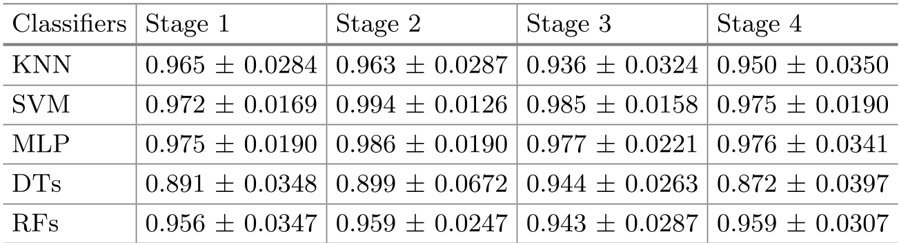 Table: Using SVM and MLP, the F1-score reached 0.994 and 0.986 respectively during the early stages of negative emotion processing.
Key observations from the data:
- Classifiers: Support Vector Machines (SVM) combined with Grid Search optimization proved robust for this small-sample, high-feature-density task.
- Valence Advantage: Negative stimuli consistently outperformed positive stimuli in discrimination tasks across almost all time windows.
- Intra vs. Inter-channel: While hemispheric asymmetry (inter-channel) is theoretically strong, intra-channel Hjorth parameters provided the most stable and highest accuracy results.
Critical Insight & Limitations
This work demonstrates that the first 1000ms of viewing a negative image reveals nearly all the information needed to identify schizophrenia-related neural anomalies.
However, there are limitations:
- Sample Size: The study used a small cohort (10 participants total). While 10-fold cross-validation helps, larger-scale validation is necessary to account for the diverse schizophrenia spectrum.
- Drug Interference: Most schizophrenia patients are on medication that can flatten EEG responses; accounting for pharmacological effects remains a hurdle for field-wide application.
Conclusion
By combining a focused emotional paradigm with efficient time-domain features, this research paves the way for a rapid, objective diagnostic tool. It shifts the focus from "what" the patient says to "how" their brain automatically reacts to the social and emotional world.