
SSMamba: Revolutionizing Pathology with Hybrid State Space Models and In-Domain SSL
SSMamba: A self-supervised hybrid state space model for pathological image classification.
SSMamba is a novel hybrid self-supervised learning (SSL) framework designed for fine-grained pathological image classification using State Space Models (SSMs). By integrating Mamba-based Masked Image Modeling (MAMIM) and domain-specific architectural modules, it achieves state-of-the-art results across 10 ROI and 6 WSI datasets, significantly outperforming 11 established pathology Foundation Models.
TL;DR
SSMamba is a task-specific framework that brings the efficiency of State Space Models (SSMs) to digital pathology. By combining a new Masked Image Modeling (MAMIM) strategy with specialized hardware-aware modules (DMS and LPR), it outperforms industry giants like Gigapath and Virchow2 on multiple benchmarks while remaining significantly more lightweight (25.3M parameters).
Background & Positioning
In the race to build the "ImageNet of Pathology," most researchers have relied on Scaling Laws: more data, more parameters, and standard Vision Transformers (ViTs). However, pathology presents unique challenges—magnification shifts, staining artifacts, and the need for both fine-grained cellular detail and global tissue structure. SSMamba positions itself as a "Specialist" rather than a "Generalist," proving that Pathology-aware architecture beats Scale-only pretraining.
Problem & Motivation: The ViT Bottleneck
Despite their success, ViT-based Foundation Models encounter high computational costs when processing high-resolution Whole Slide Images (WSIs). Furthermore:
- Domain Shift: Models trained at 20x magnification often fail when applied to 40x or 10x clinical images.
- Local-Global Duality: Standard attention mechanisms are great at global context but often "blur" the fine-grained cellular boundaries (e.g., nuclear-cytoplasmic interfaces) essential for grading cancer.
- Translation Invariance: Traditional positional encodings are sensitive to the random rotations and shifts common in tissue slicing.
Methodology: The Core of SSMamba
SSMamba introduces three architectural breakthroughs to solve these issues:
1. MAMIM (Mamba Masked Image Modeling)
Unlike standard MAE which uses ViT blocks, MAMIM uses SSM-based blocks to reconstruct 75% masked patches. This forces the model to learn the underlying "grammar" of tissue topology and cell morphology.
2. DMS (Directional Multi-scale) Module
Vanilla Mamba is unidirectional (1D sequence). DMS transforms this for 2D pathology by:
- Bidirectional Scanning: Replacing causal convolutions with bidirectional depthwise separable convolutions to capture spatial context from all sides.
- Parallel Convolutional Branch: Adding a symmetric CNN branch to extract local cellular patterns that linear SSM scans might miss.
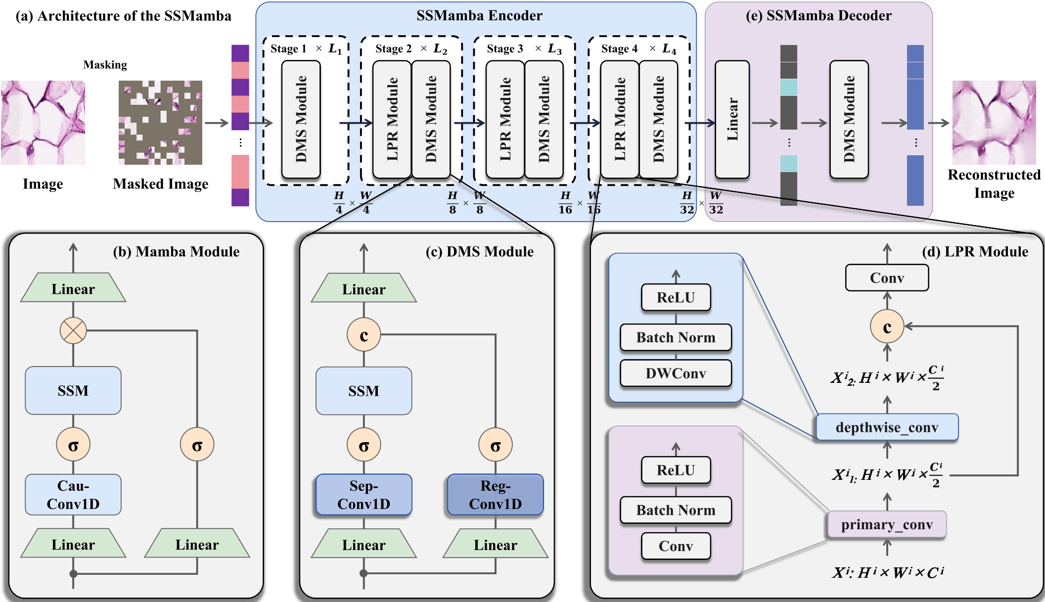 Figure 1: The hierarchical SSMamba encoder featuring the DMS and LPR modules.
3. LPR (Local Perception Residual) Module
To replace rigid positional embeddings, LPR uses depthwise convolutions (DWConv) and residual connections. This makes the model translation-invariant and robust to staining variations (H&E artifacts) by focusing on relative local structures rather than absolute coordinates.
Experiments & Results: David vs. Goliath
The experimental results are striking. SSMamba, with only 25.3 million parameters, frequently outperformed billion-parameter models.
- SOTA Benchmarking: On the CAM16 dataset, SSMamba achieved 93.51% Accuracy, significantly higher than the massive Gigapath (88.42%).
- Efficiency: As shown in the "Bubble Chart" below, SSMamba occupies the "sweet spot" of high performance and low parameter count.
- Cross-Dataset Generalization: When trained on one dataset (NCT) and tested on others (MHIST/CRC), SSMamba maintained a high F1-score (~87%), whereas competitive models like MUSK saw significant performance drops due to domain shift.
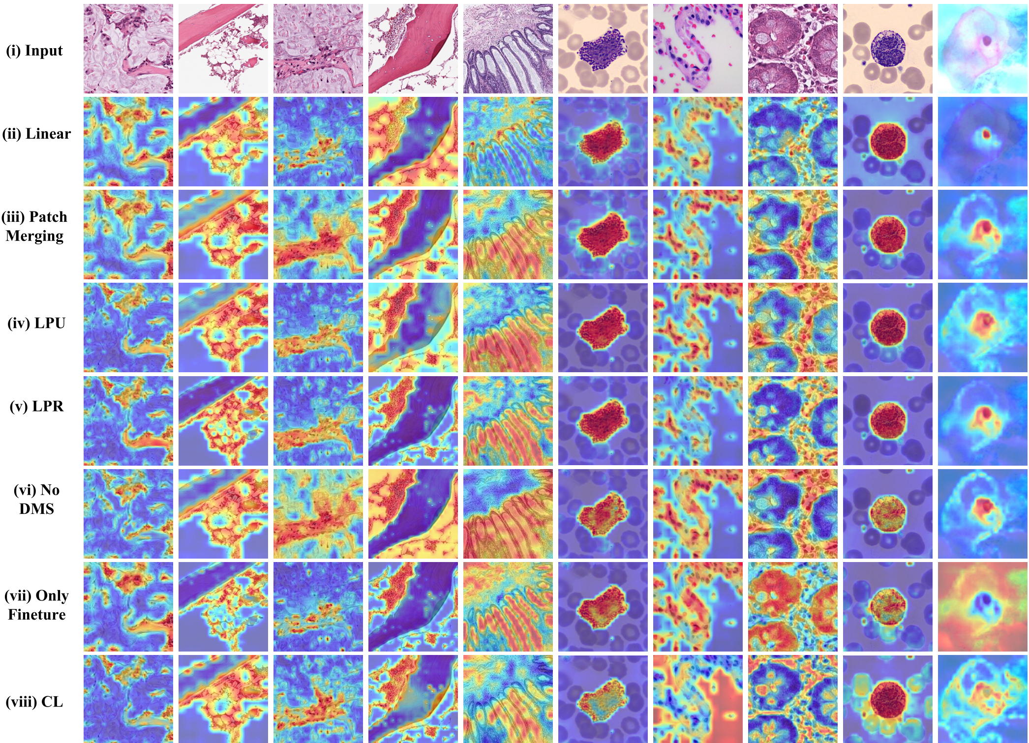 Figure 2: Performance vs. Parameter Count. SSMamba achieves SOTA accuracy with minimal resource consumption.
Visual Evidence: Grad-CAM Analysis
Visualizations using Grad-CAM confirm that SSMamba "looks" at the right things. While ViT results are often over-homogenized and VMamba shows "striping" artifacts, SSMamba identifies sharp cellular boundaries and critical tumor-stroma interfaces.
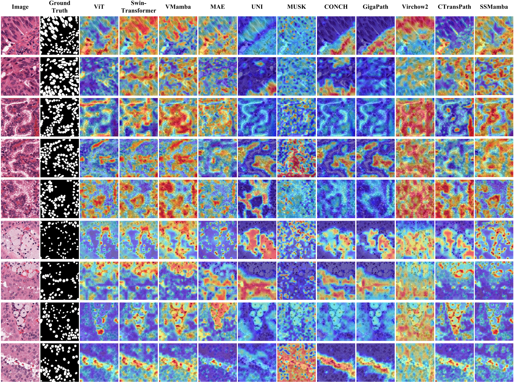 Figure 3: Grad-CAM comparison showing SSMamba's precise localization of diagnostic regions.
Critical Analysis & Conclusion
Takeaway
SSMamba proves that the Mamba architecture is naturally suited for pathology. Its linear complexity solves the resolution problem of WSIs, while the DMS and LPR modules provide the necessary inductive biases for fine-grained medical analysis.
Limitations
Currently, the framework is optimized for classification. While the authors demonstrated success on both ROI and WSI levels, its performance in dense tasks like segmentation (masking every cell) or mitosis detection remains to be fully explored in future work.
Future Outlook
The success of SSMamba suggests that the future of Medical AI may not lie in bigger models, but in smarter architectures that respect the biological and physical properties of the data they process.